Page 1
23
Neural Images
Raymond Tak-Fai Cheung
Case 23.1
Leading question
A 55-year-old woman with a history of hypertension presents with
sudden onset of unilateral weakness.
View original page (figures / layout)
Page 2
354 Problem-Based Medical Case Management Case 23.1
Questions
1.
2.
3.
4.
Describe two CT abnormalities.
‘What is the CT diagnosis?
Name two neurological deficits likely present.
‘What is the most likely underlying aetiology?
Answers
1.
2.
Left subcortical/lenticular/basal ganglial hyperdense lesion;
mass effect/obliteration of left lateral ventricle/midline shift.
Acute left intracerebral haemorrhage/subcortical haematoma/
lentiform haematoma.
Right hemiparesis/hemiplegia, right-sided facial weakness of
upper motor neurone pattern, dysarthria, right hemisensory
loss.
Hypertension.
Description
Hyperdense lesions in plain CT of the head are due to haemor-
rhage or calcification.
Calcification is typically symmetrical and not space-occupying.
Haematoma is space-occupying and leads to obliteration of
ventricles, effacement of sulcal spaces, and midline shift.
Intracerebral haemorrhage typically produces contralateral
weakness of limbs and face, contralateral sensory loss over
limbs and face, and dysarthria.
Discussion
About 20-25% of strokes are due to intracerebral haemorrhage.
Common causes include hypertension, bleeding tendency, and -
cerebral amyloid angiopathy.
Less common causes include arteriovenous malformation, cav-
ernoma, thrombolysis, aneurysm, Moyamoya disease, tumour,
vasculitis, and drug abuse. .
View original page (figures / layout)
Page 3
Case 23.2 Neural Images 355
+ Typical locations of hypertensive intracerebral haemorrhage
include lentiform nucleus, thalamus, brainstem, and cerebellum.
« Medical or conservative management is usually adopted for
deep-seated haemorrhage. Neurosurgical evacuation may be
life-saving, but does not improve the quality of life.
Case 23.2
Leading question
A 53-year-old man with a history of hypertension, diabetes mel-
litus, hypercholesterolaemia, and atrial fibrillation presents with
sudden onset of unilateral weakness and language deficits.
View original page (figures / layout)
Page 4
356 Problem-Based Medical Case Management Case 23.2
Questions
1.
Describe the CT abnormalities.
2. What is the CT diagnosis?
3. Name three neurological deficits likely present.
4. What preventive measures should be considered?
Answers
1.
Left cortical and subcortical hypodense lesion involving the
lentiform nucleus, caudate nucleus, frontal and temporal
cortex (left middle cerebral artery territory); presence of some
isodense areas within the hypodense lesion due to petechial
haemorrhagic transformation.
. Recent left middle cerebral artery territory ischaemic infarc-
tion/stroke with petechial haemorrhagic transformation of
infarction.
Right hemiparesis/hemiplegia, right-sided facial weakness of
upper motor neurone pattern, aphasia, dysarthria, right hemi-
sensory loss.
Optimal control of hypertension and diabetes mellitus, use of
aspirin or another antiplatelet agent, use of a statin, consider-
ing long-term anti-coagulation with warfarin or a new oral
anti-coagulant.
Description
Hypodense lesions in plain CT of the head are due to infarction
or oedema.
Oedema mainly involves the subcortical white matter, produc-
ing a finger-like pattern.
Infarction involves both grey and white matter unless it is small.
Haemorrhagic transformation occurs upon spontaneous Or
therapeutic reperfusion, leading to petechial haemorrhage or
frank haematoma.
View original page (figures / layout)
Page 5
Case 23.3 Neural Images 357
Left middle cerebral artery infarction produces right-sided
weakness over limbs and face, right-sided sensory loss over
limbs and face, aphasia, dysarthria.
Discussion
About 70-75% of strokes are due to cerebral infarction (ischae-
mic stroke).
Common causes include atherothrombosis, thromboembolism,
cardioembolism, and small vessel arteriolosclerosis.
Less common causes include dissection, hypercoagulopathy,
Moyamoya disease, vasospasm, vasculitis, drug abuse, and
migraine.
Common risk factors are hypertension, diabetes mellitus, hyper-
cholesterolaemia, extra- or intracranial stenosis, atrial fibril-
lation, valvular heart disease, ischaemic heart disease, other
cardioembolic conditions, smoking, alcohol abuse, obesity,
advanced age, and male gender.
Preventive measures include identification and optimal con-
trol of all modifiable risk factors, use of aspirin or another
antiplatelet agent, surgical or intervention for severe extracra-
nial stenosis, considering anti-coagulation for cardioembolic
strokes, cessation of smoking, moderation of drinking, regular
exercise, and weight reduction.
Case 23.3
Leading question
A 75-year-old woman with good past health presents with sudden
onset of unilateral weakness. (See p. 358.)
View original page (figures / layout)
Page 6
358 Problem-Based Medical Case Management Case 23.3
Questions
1. Describe the CT abnormalities.
2. What is the CT diagnosis?
3. Please name two neurological deficits likely present.
4. What is the most likely underlying aetiology?
Answers
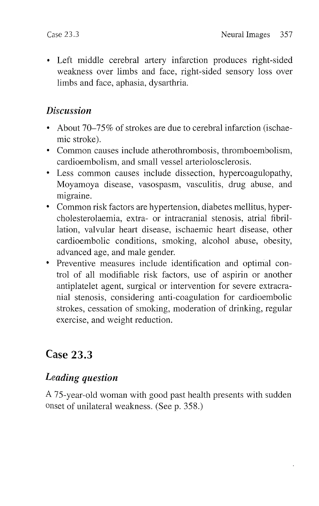
1. Right frontoparietal hyperdense lesion with surrounding
hypodense rim; sulcal effacement from mass effect.
2. Acute right lobar intracerebral haemorrhage.
3. Left hemiparesis/hemiplegia, left-sided facial weakness of
upper motor neurone pattern, dysarthria, left-sided cortical
sensory loss.
4. Cerebral amyloid angiopathy.
View original page (figures / layout)
Page 7
Case 23.4 Neural Images 359
Description
.
Hyperdense lesions in plain CT of the head are due to haemor-
rhage or calcification.
Calcification is typically symmetrical and not space-occupying.
Haematoma is space-occupying and leads to obliteration of
ventricles, effacement of sulcal spaces, and midline shift.
Intracerebral haemorrhage typically produces contralateral
weakness of limbs and face, contralateral sensory loss over
limbs and face and dysarthria. Involvement of parietal cortex
may cause contralateral cortical sensory loss.
Discussion
About 20-25% of strokes are due to intracerebral haemorrhage.
Common causes include hypertension, bleeding tendency, and
cerebral amyloid angiopathy. Patients with cerebral amyloid
angiopathy are usually 70 years or older.
Less common causes include arteriovenous malformation, cav-
ernoma, thrombolysis, aneurysm, Moyamoya disease, tumour,
vasculitis, and drug abuse.
Typical locations of intracerebral haemorrhage from cerebral
amyloid angiopathy are lobar, subarachnoid, and mutifocal.
Recurrent haemorrhage is common.
Medical or conservative management is usually adopted.
Neurosurgical biopsy may confirm amyloid angiopathy, but
haemostasis is a concern.
Case 23.4
Leading question
A 45-year-old man with good past health presents with sudden
onset of unilateral facial numbness and limb ataxia, contralateral
numbness, dysphagia, and vertigo. He receivad vigorous massage
over his neck 3 days ago. (See p. 360.)
View original page (figures / layout)
Page 8
360 Problem-Based Medical Case Management Case 23.4
Questions
1. Describe the abnormalities.
2. What is the diagnosis?
3. Describe the neurological deficits.
4. What is the most likely underlying aetiology?
Answers
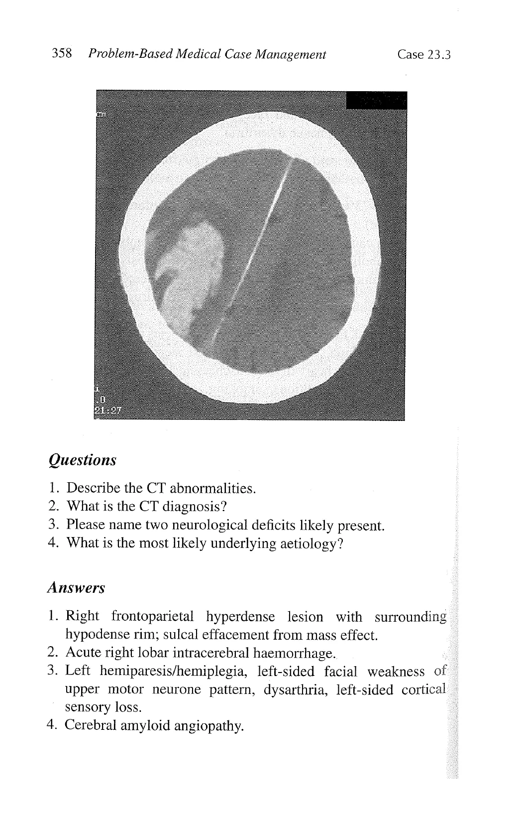
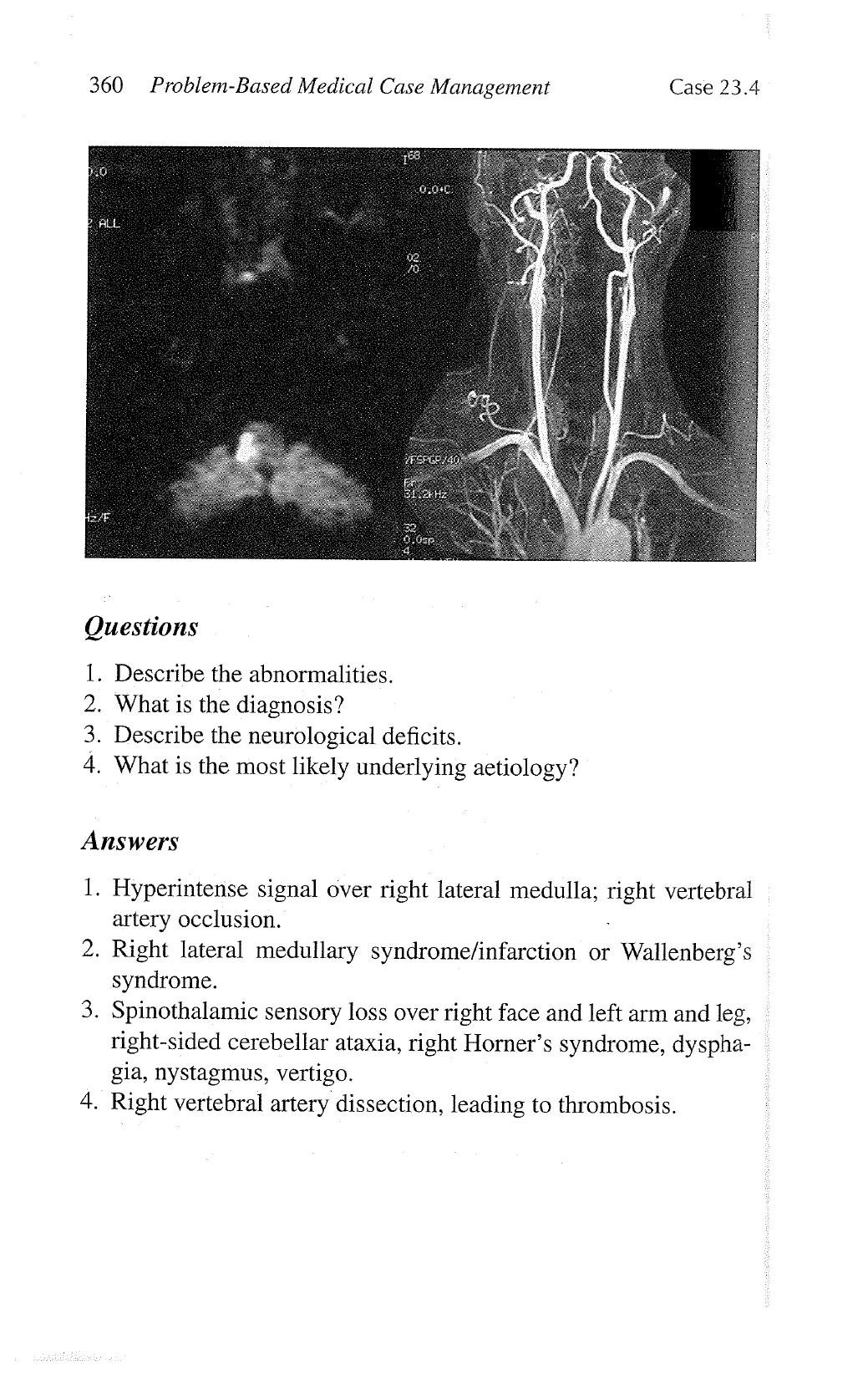
1. Hyperintense signal over right lateral medulla; right vertebral
artery occlusion.
2. Right lateral medullary syndrome/infarction or Wallenberg’s
syndrome.
3. Spinothalamic sensory loss over right face and left arm and leg,
right-sided cerebellar ataxia, right Horner’s syndrome, dyspha-
gia, nystagmus, vertigo.
4. Right vertebral artery dissection, leading to thrombosis.
View original page (figures / layout)
Page 9
Case 23.4 Neural Images 361
Description
Diffusion-weighted imaging reveals the acute infarction in the
form of a hyperintense signal.
Magnetic resonance angiography shows the luminal anatomy of
cerebral arteries.
Lateral medullary infarction can result from thrombosis of ver-
tebral artery, basilar artery, posterior inferior cerebellar artery,
anterior inferior cerebellar artery or superior cerebellar artery.
There is involvement of trigeminal nucleus and tract, spinocer-
ebellar tract, spinothalamic tract, nucleus solitarius, nucleus
ambiguus, ninth and tenth nuclei, and sympathetic fibres.
Discussion
Arterial dissection should be considered in young stroke patients
without vascular risk factors especially if there is a history of
recent trauma or manipulation of the neck.
Tears occur in a major neck artery with bleeding into the arterial
wall, leading to stenosis, occlusion, and/or aneurismal dilata-
tion. Spontaneous dissection also oceurs, especially in patients
with Ehlers-Danlos syndrome, Marfan syndrome, polycystic
kidney disease, osteogenesis imperfecta type I, fibromuscular
dysplasia, and cystic medial necrosis.
Dissection can be asymptomatic. Characteristic features include
unilateral headache, facial or neck pain, Horner’s syndrome,
recurrent transient ischaemic attacks, cranial nerve palsies, pul-
satile tinnitus, and audible bruit.
Dissection can be confirmed by digital subtraction angiography,
magnetic resonance angiography, and CT angiography.
Anti-coagulation for 3-6 months is considered. Aspirin is an
alternative and also for long-term use if luminal irregulari-
ties persist. Endovascular or surgical treatment is reserved for
patients with persistent symptoms despite anti-coagulation.
View original page (figures / layout)
Page 10
362 Problem-Based Medical Case Management Case 23.5
Case 23.5
Leading question
A 36-year-old man with good past health presents with confused
speech, poor memory, and disorientation. He has had flu-like
illness for 3 days.
Questions
1. Describe the magnetic resonance imaging abnormalities.
2. What is the diagnosis?
3. Name the possible complications.
4. Outline the specific treatment.
View original page (figures / layout)
Page 11
Case 23.5 Neural Images 363
Answers
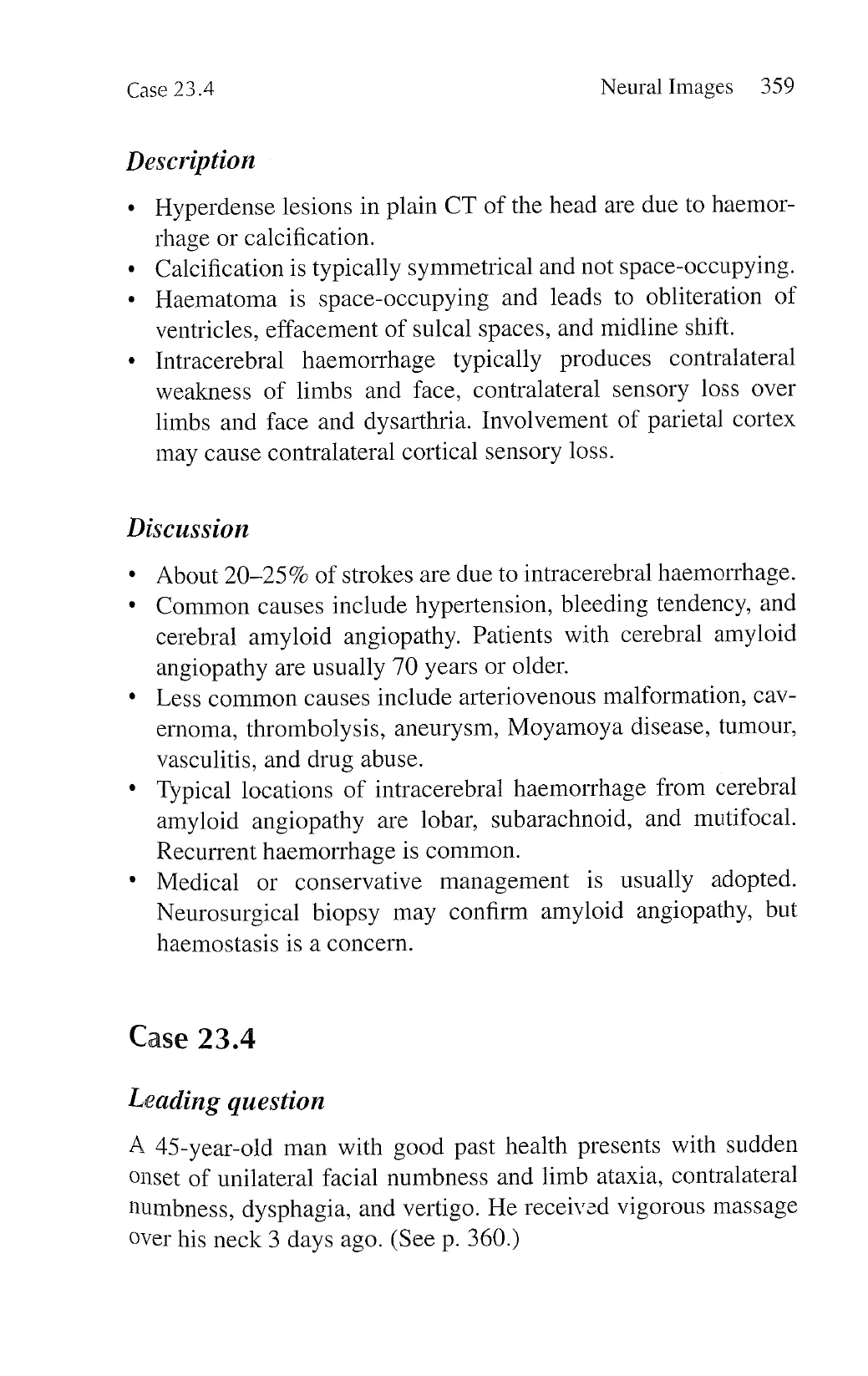
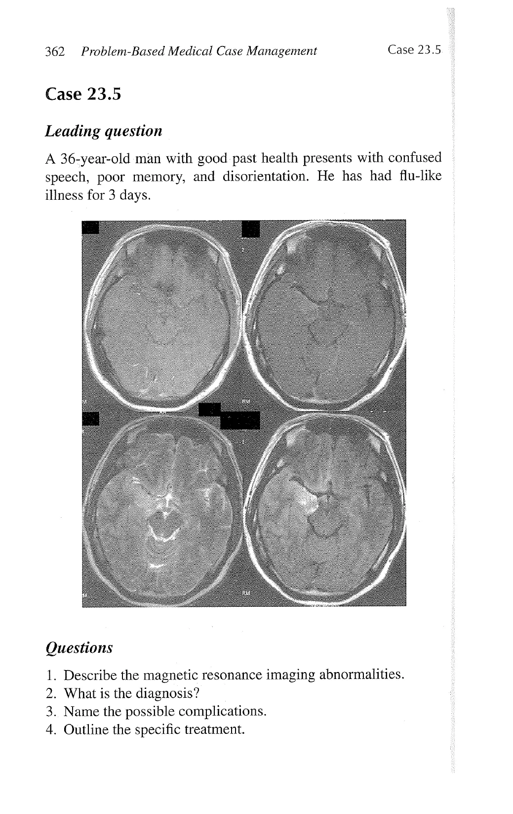
1. Hyperintense signal over right mesiotemporal region with
mildly thickened cortices over the right temporal lobe in
the T2-weighted and FLAIR images; abnormal gadolinium
enhancement over the right mesiotemporal region in the
T1-weighted image.
Herpes simplex encephalitis (viral encephalitis with temporal
lobe involvement).
3. Epileptic seizures, aphasia, hemiparesis, amnesia, coma.
4. Acyclovir, anticonvulsant, corticosteroids.
IS
Description
* Hyperintense signal on T2-weighted and FLAIR images indi-
cates increased water content. Abnormal enhancement with
gadolinium indicates break-down of the blood-brain barrier.
* Predominant involvement of cortex suggests encephalitis.
* Herpes simplex encephalitis has a predilection for the temporal
and frontal lobes.
Discussion
* Encephalitis refers to inflammation of brain tissue. Grey matter
is predominately involved in acute viral encephalitis; coexisting
viral meningitis is common. Post-infectious encephalitis typi-
cally causes white matter demyelination.
* Features include non-specific symptoms of infection, altered
mental state, seizures, focal deficits, aphasia, amnesia, and
myoclonus.
* CT may be normal. Useful investigations include magnetic res-
onance imaging, cerebrospinal fluid analyses, polymerase chain
reaction, paired sera for viral titre, and electroencephalogram.
Brain biopsy is seldom performed nowadays.
* Herpes simplex type 1 is the most common cause of sporadic
encephalitis worldwide. There is a predilection for the temporal
and frontal lobes.
View original page (figures / layout)
Page 12
364 Problem-Based Medical Case Management Case 23.6
* Specific treatment includes acyclovir, anticonvulsant, and corti-
costeroids. Mortality and disability are high if left untreated or
treatment is delayed.
Case 23.6
Leading question
A 19-year-old college student with good past health presents
with low-grade fever, headache, nausea, vomiting, and ataxia.
Cerebrospinal fluid examination shows a total cell count of 120
per puL (86% lymphocytes), protein concentration of 2.6 g/L, and
glucose concentration of 1.8 mmol/L (plasma glucose level = 5.8).
Questions
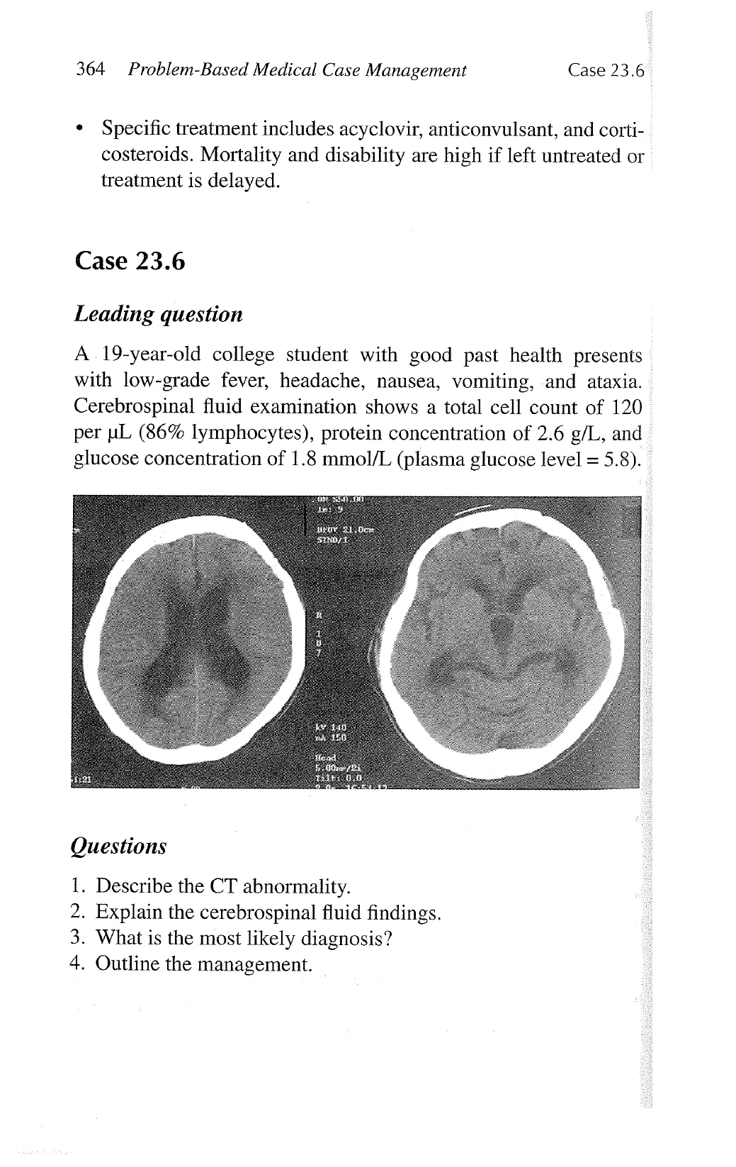
1. Describe the CT abnormality.
2. Explain the cerebrospinal fluid findings.
3. What is the most likely diagnosis?
4. Outline the management.
View original page (figures / layout)
Page 13
Case 23.6 Neural Images 365
Answers
1.
Dilated lateral and third ventricles with sulcal effacement
indicative of obstructive hydrocephalus.
Lymphocytic pleocytosis with low glucose level and high
protein level indicative of chronic bacterial meningitis (e.g.,
tuberculosis) or fungal meningitis.
Tuberculous meningitis with obstructive hydrocephalus.
. Multiple anti-tuberculous drugs for 9-12 months, shunting
of hydrocephalus, a course of corticosteroids, monitoring
of response with clinical and radiological assessment, and
repeated lumbar puncture.
Description
Ventricles are slit-like in young subjects. Dilated ventricles
with sulcal effacement suggest obstructive hydrocephalus.
Although lymphocytic pleocytosis is consistent with viral men-
ingitis, low glucose level is against this possibility. High protein
level indicates a chronic type of infection.
Urgent external ventricular drainage and/or ventriculo-perito-
neal shunting should be considered in obstructive hydrocepha-
lus. Lumbar puncture is contra-indicated.
Extra-pulmonary tuberculosis requires a longer duration of
standard anti-tuberculous chemotherapy. A course of corti-
costeroids is indicated in stage II or I tuberculous meningitis
(lethargy, prominent meningeal irritation, cranial nerve palsies,
hydrocephalus, convulsion, paralysis, stupor or coma).
Discussion
.
Acute rather than chronic presentation of tuberculous meningi-
tis is common in Hong Kong.
Possible complications include hydrocephalus, cranial nerve
palsies, tuberculoma, spinal block, stroke, tuberculous brain
abscess, and myeloradiculopathy.
View original page (figures / layout)
Page 14
366 Problem-Based Medical Case Management Case 23.7
» Delayed treatment is associated with a high mortality and
disability.
* Anti-tuberculous chemotherapy carries a high risk of adverse
effects: isoniazid (hepatitis, peripheral neuropathy); rifampicin
(drug interactions, hepatitis, fever and vasculitis, nausea and
vomiting); ethambutol (optic neuritis, hepatitis, peripheral neu-
ropathy); pyrazinamide (hepatitis, gout).
Case 23.7
Leading question
A 52-year-old man presents with gradual onset of headache,
nausea, and vomiting. He also has several episodes of involuntary
twitching of his right leg. His serum and urine osmolality are 250
and 500 milliosmole/kg, respectively. He is a chronic smoker.
View original page (figures / layout)
Page 15
Case 23.7 Neural Images 367
Questions
W —
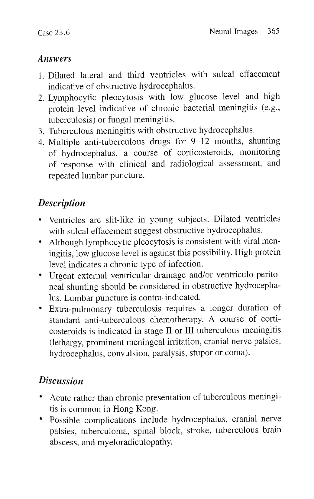
. Describe the abnormalities.
. What is the most likely diagnosis?
. What complications have developed?
. Outline the management for his neurological condition and the
above complications.
Answers
L.
Hypodense lesions in the form of finger-like projections over
the left frontal and parietal regions as well as the right frontal
and parieto-occipital regions; contrast-enhancing nodules over
the right fronto-parietal regions; sulcal effacement, obliteration
of lateral ventricle, and right-to-left midline shift indicative of
mass effects.
. Cerebral metastases (likely to have underlying carcinoma of
lung).
Focal motor epileptic seizures, raised intracranial pressure, syn-
drome of inappropriate antidiuretic hormone secretion.
High-dose corticosteroids for vasogenic oedema and raised
intracranial pressure, prophylactic anti-convulsant for focal
seizures, palliative whole brain irradiation for cerebral metas-
tases and fluid restriction plus sodium chloride supplement for
syndrome of inappropriate antidiuretic hormone secretion.
Description
Hypodensity on CT indicates cytotoxic or vasogenic oedema.
Vasogenic oedema predominately affects the white matter,
leading to a finger-like projection pattern.
Sulcal effacement, obliteration of ventricles, and midline shift
indicate mass effect.
View original page (figures / layout)
Page 16
368 Problem-Based Medical Case Management Case 23.8
Contrast-enhancement indicates breakdown of blood-brain
barrier, e.g., metastasis, inflammation, infection, infarction.
Low serum osmolality and high urine osmolality indicate inap-
propriate secretion of antidiuretic hormone.
Episodic involuntary contractions of a limb suggest focal motor
seizures.
Discussion
Cerebral metastasis commonly arises from carcinomas of lung,
breast, kidney, and colon as well as from choriocarcinoma and
malignant melanoma.
Secondaries to the brain are more common than primary brain
tumours.
Focal features of insidious onset and gradual progression are
typical. Focal seizures and raised intracranial pressure are
common.
CT or magnetic resonance imaging of the brain with and
without contrast, electroencephalogram, and screening of the
primary are appropriate. Lumbar puncture is contra-indicated.
Case 23.8
Leading question
A 62-year-old man with hypertension, diabetes mellitus, and
hypercholesterolaemia presents with an episode of transient blind-
ness of his left eye. He is a chronic smoker and a heavy drinker.
Neck bruits are present.
View original page (figures / layout)
Page 17
Case 23.8 Neural Images 369
Questions
1. Describe the abnormality.
2. What is the diagnosis of his transient neurological symptom?
3. What complication is he at risk of developing?
4. Qutline the treatment options.
Answers
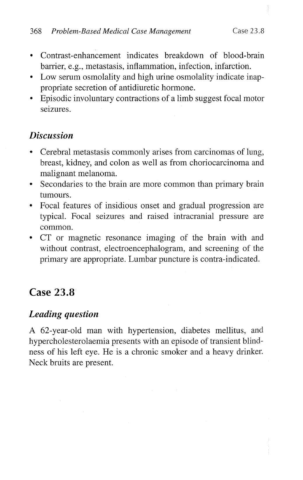
1. Severe stenosis (narrowing) of his proximal left internal carotid
artery.
2. Left amaurosis fugax (transient monocular blindness).
. Left middle cerebral artery territory ischaemic stroke.
4. Control of risk factors, use of an antiplatelet agent, carotid
endarterectomy or percutaneous transluminal angioplasty and
stenting, cessation of smoking, and moderation of drinking.
W
View original page (figures / layout)
Page 18
370 Problem-Based Medical Case Management Case 23.8
Description
Digital subtraction angiography is the gold-standard examina-
tion for suspected arterial lesions.
Severe stenosis of the internal carotid artery is associated with a
higher risk of ipsilateral transient ischaemic attacks or strokes.
Mechanisms include thromboembolism and haemodynamic
compromise.
The ophthalmic artery is a branch of the internal carotid artery.
The internal carotid artery terminates into the middle cerebral
artery.
Discussion
Internal carotid artery stenosis is an important cause of ischae-
mic stroke, especially in Caucasians.
The actual risk of stroke is increased by the following factors:
occurrence of symptoms, higher degree of stenosis, presence
of ulceration, contralateral occlusion, inadequate collaterals,
coexistence of multiple risk factors, and cerebral infarction
on CT.
Use of aspirin or another antiplatelet agent and control of risk
factors should be adopted in all patients.
Carotid endarterectomy carries a risk of stroke, death, cranial
nerve palsy, hoarseness of voice, and wound complications.
Carotid endarterectomy is indicated in patients with severe
symptomatic stenosis of the internal carotid artery if the risk of
surgery is less than 6%.
Percutaneous transluminal angioplasty and stenting are
alternatives.
View original page (figures / layout)
Page 19
Case 23.9 Neural Images 371
Case 23.9
Leading question
A 60-year-old woman with good past health presents with sudden
onset of severe occipital headache, neck stiffness, and vomiting.
Questions
1. Describe the abnormality.
2. What is the diagnosis?
3. What are the useful investigations?
4. What other neurological features are possible?
5. Outline the management.
View original page (figures / layout)
Page 20
372 Problem-Based Medical Case Management Case 23.9
Answers
1.
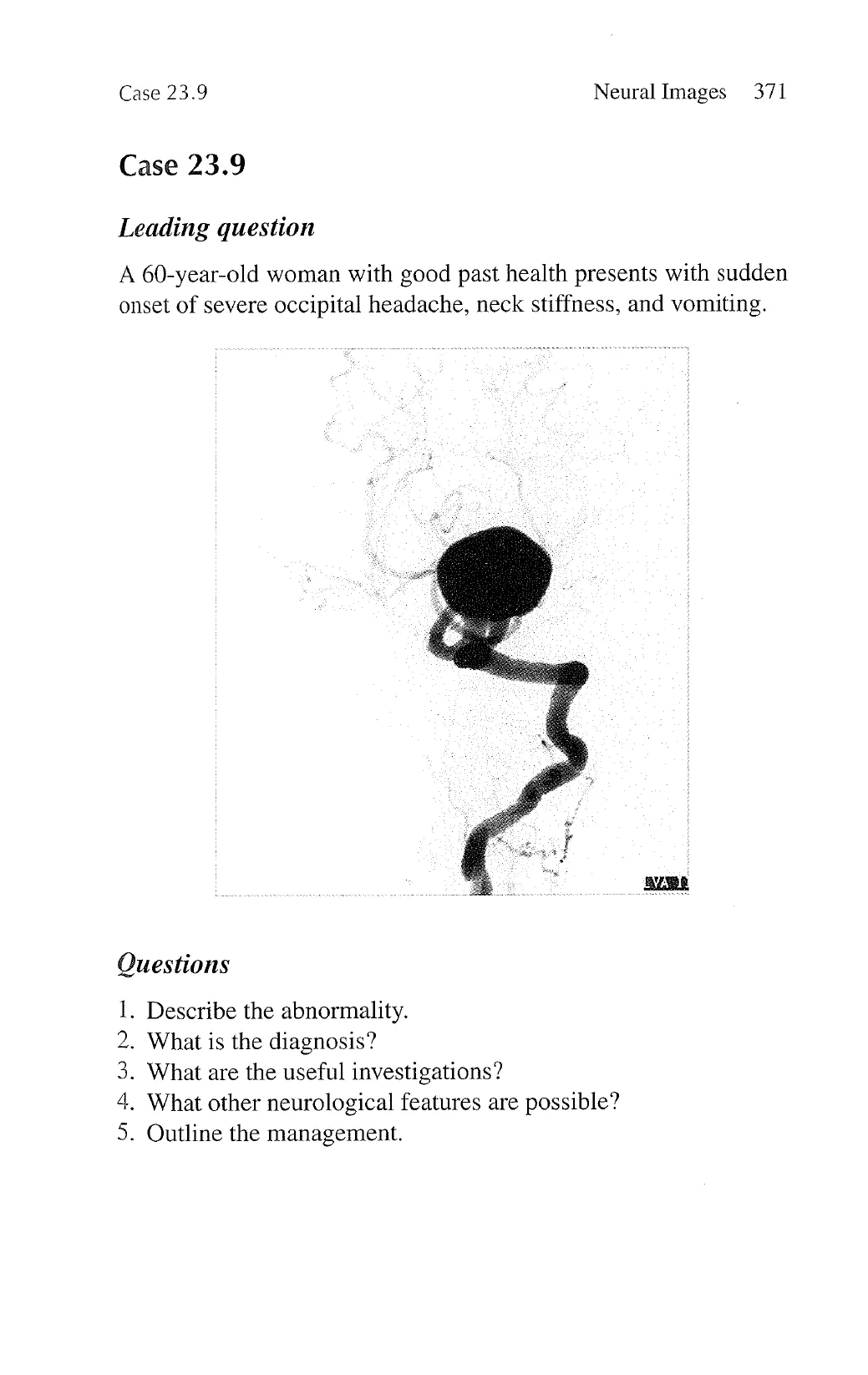
. Subarachnoid haemorrhage from leakage of the giant aneurysm.
. CT of the head, lumbar puncture, and cerebrospinal fluid
A giant fusiform aneurysm of the basilar artery.
analysis.
. Features of brainstem compression, thromboembolic events to
brainstem and posterior cerebral artery territory (homonymous
hemianopia, thalamic sensory symptoms).
Neurosurgical clipping and decompression or endovascular
coiling of the aneurysm, prophylactic anti-convulsant, close
neuro-observation, surveillance of complications, a course of
calcium antagonist (e.g., nimodipine).
Description
Digital subtraction angiography is the gold-standard examina-
tion for suspected arterial lesions.
Abnormal dilatation of an artery is termed aneurysm. The shape
can be fusiform or saccular.
An aneurysm may rupture into subarachnoid space to cause
subarachnoid haemorrhage or into brain parenchyma to cause
intracerebral haemorrhage.
A large aneurysm has a greater risk of rupture and may com-
press onto the neighboring structures. Turbulent flow inside
may lead to thromboembolic events.
Discussion
Typical features of subarachnoid haemorrhage are severe head-
ache of sudden onset, neck stiffness, and symptoms of raised
intracranial pressure. Impaired consciousness is common.
CT shows hyperdense signal of the blood in the subarachnoid
space, ventricles, and/or cisterns in 95% of patients.
Presence of blood in cerebrospinal fluid is also diagnostic.
Lumbar puncture is usually reserved for CT-negative cases.
Examination for xanthochromia is crucial when there is a
View original page (figures / layout)
Page 21
Case 23.10 Neural Images 373
relatively long interval between the suspected bleeding and
[umbar puncture.
» Complications include rebleeding, vasospasm, hydrocephalus,
cerebral oedema, seizure, syndrome of inappropriate anti-
diuretic hormone secretion, cardiac arrhythmias, and other
complications.
Case 23.10
Leading question
A 55-year-old man with hypertension and diabetes mellitus pre-
sents with a 2-year history of episodic neck pain with radiation
down to the arms, a 6-month history of bilateral arm weakness and
a 3-month history of difficulty in walking.
View original page (figures / layout)
Page 22
374 Problem-Based Medical Case Management Case 23.10
Questions
1.
Describe the abnormalities.
2. What is the diagnosis?
3.
Describe the neurological deficits.
4. Outline the treatment options.
Answers
1.
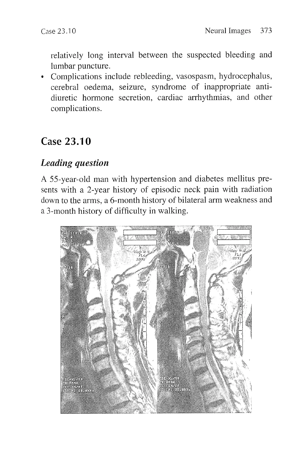
Spondylotic changes of the cervical spine; obliteration of the
cerebrospinal space anterior and posterior to the cervical spinal
cord from herniating intervertebral discs; osteophytes, ossified
posterior longitudinal ligament, and hypertrophied ligamentum
flavam; compression of the cervical spinal cord at multiple
levels.
Cervical spondylotic radiculomyelopathy.
. Cervical radiculopathy with sensory symptoms and lower-
motor-neurone pattern of weakness over multiple cervical
roots, spastic paraparesis, and dorsal column sensory deficits
from cervical myelopathy due to compression of the lateral and
dorsal columns.
Conservative management with traction, physiotherapy, neck
collar, and analgesics; surgical decompression with anterior
spinal fusion.
Description
Water is hyperintense on T2-weighted magnetic resonance
imaging.
Presence of ample cerebrospinal fluid-filled space both anterior
and posterior to the cord is normal.
Hyperintense signal within the cord may be present to indicate
cord oedema.
View original page (figures / layout)
Page 23
Case 23.10 Neural Images 375
Discussion
Degenerative tissues of the spine may compress the spinal cord
and roots. These tissues include osteophytes, herniating discs,
ossified posterior longitudinal ligament, and hypertrophied
ligamentum flavum.
Cervical spondylosis is a very common radiological diagnosis,
and cervical spondylotic radiculomyelopathy is a common
cause of cervical cord and root lesion in people aged 50 or
older.
Pain over the neck with radiation down the upper limb dermato-
mes is common. Compression of cervical roots leads to lower-
motor-neurone motor deficits and sensory symptoms over the
arms. Cervical cord compression leads to upper-motor-neurone
motor deficits over the legs. Dorsal column sensory loss over
the legs may be present.
Magnetic resonance imaging and somatosensory evoked poten-
tial study are useful investigations.
Conservative management is indicated in patients with mild
stable symptoms. Surgical treatment is indicated in patients
with significant symptoms or progression and for those who fail
conservative treatment.
View original page (figures / layout)