Page 1
24
Plain X-Ray Images
Chi-Ho Lee and Chak-Sing Lau
Case 24.1
Leading question
A 45-year-old man presents with abdominal pain and frequent
bumping into corners at home.
View original page (figures / layout)
Page 2
Case 24.1 Plain X-Ray Images 377
Questions
1 —
o
Name one abnormality on the skull X-ray.
. What further imaging could confirm the abnormality?
. What causes the abdominal pain?
. Explain the problem of frequent bumping into corners.
. What is the clinical syndrome?
Answers
W -
v
Double flooring of pituitary fossa.
Magnetic resonance imaging (MRI) of the pituitary.
Renal colic.
Visual field defect.
Multiple endocrine neoplasia Type 1.
Description
Expansion of the sella turcica.
Enlarged frontal sinus.
Thickened skull vault.
Prognathous jaw.
There is ballooning of the pituitary fossa, erosion and destruc-
tion of the anterior, posterior clinoids, and of the tuberculum
sellae. Enlargement of paranasal sinuses and widening of the
mandibular angle with prognathism.
Discussion
Space-occupying lesions of the pituitary gland produce distur-
bances in two aspects: structural and functional.
Structural: space-occupying lesion of the pituitary fossa if
larger than 1 cm can compress on the optic chiasm, producing
bitemporal hemianopia.
View original page (figures / layout)
Page 3
378 Problem-Based Medical Case Management Case 24.1
e In this patient with acromegaly, the pituitary fossa is greatly
enlarged due to the directly expanding effect of an adenoma
of the anterior pituitary gland. A “double flooring” of the sella
occurs when the tumour expands downwards unevenly. His
MRI pituitary shows a pituitary macroadenoma.
Coronal image Saggital image
« Functional: hormonal disturbances originating from the pitui-
tary gland involve either hyper- or hypo-secretion of hormones.
« Deficiency of ACTH, TSH or antidiuretic hormone, alone or in
combination may lead to life-threatening situations.
* Hormonal hypersecretion is usually due to hormone-secreting
pituitary tumours, notably growth hormone-secreting (acro-
megaly), prolactinoma, and ACTH-secreting (Cushing’s
disease) tumours.
Acidophilic or chromophobe adenomas of the pituitary are
occasionally associated with functioning islet cell tumours of
the pancreas, and adenomas of the parathyroids giving rise to
the disorder, called multiple endocrine neoplasia Type 1. The
disorder is frequently familial and may present with symptoms
of the pituitary tumour, hypoglycaemia, hypercalcaemia or gas-
trointestinal bleeding.
¢ Renal colic is due to renal stones secondary to hypercalcaemia.
View original page (figures / layout)
Page 4
Case 24.2 Plain X-Ray Images 379
Case 24.2
Leading question
A 65-year-old woman presented to her general practitioner with
acute back pain located at the upper lumbar region. X-rays of the
lumbar spine were taken.
Ibuprofen 400 mg t.i.d. was prescribed, but there was only
partial improvement in the patient’s back pain.
Questions
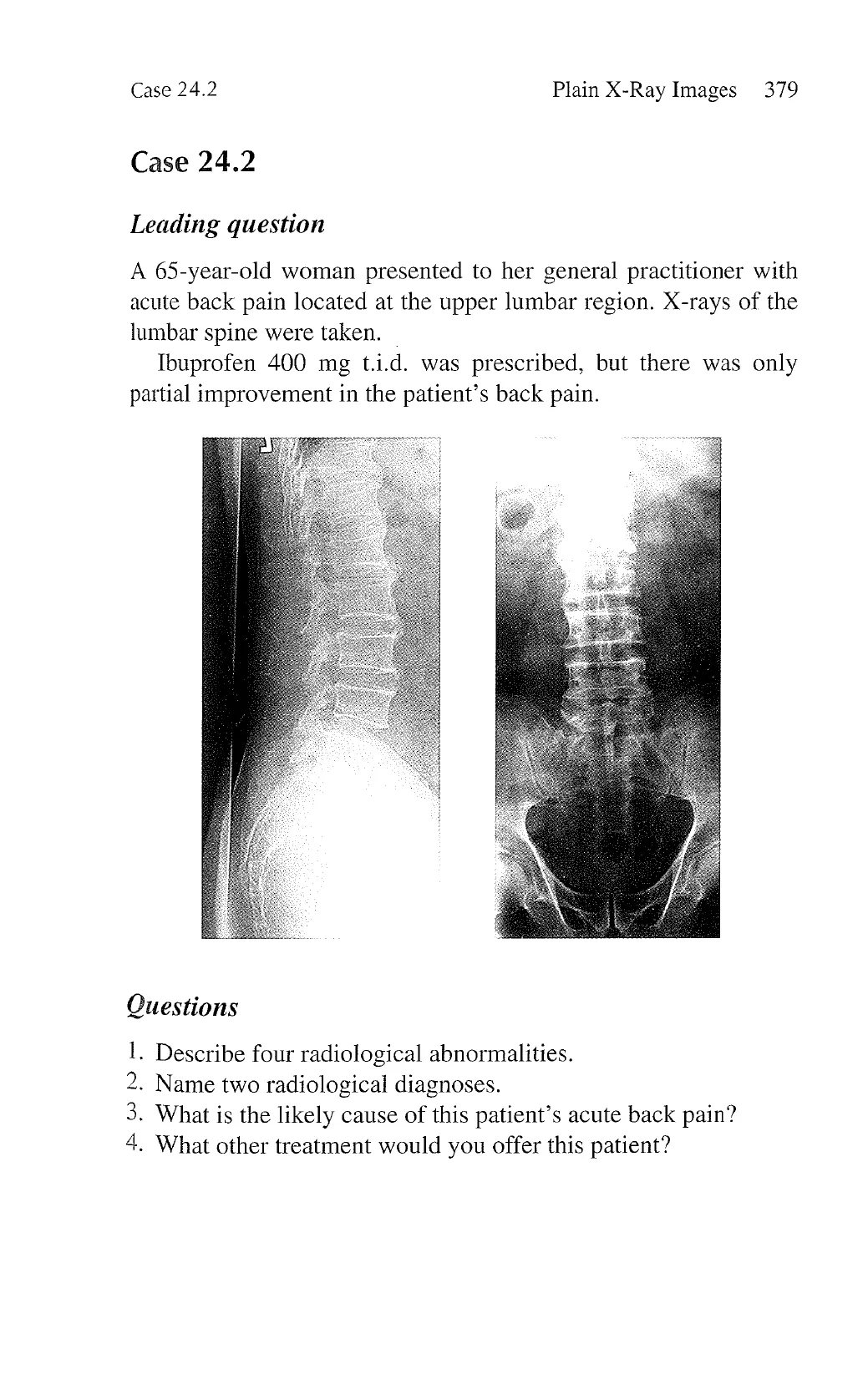
1. Describe four radiological abnormalities.
2. Name two radiological diagnoses.
3. What is the likely cause of this patient’s acute back pain?
4. What other treatment would you offer this patient?
View original page (figures / layout)
Page 5
380 Problem-Based Medical Case Management Case 24.2
Answers
1.
Y
Loss of joint space between L5 vertebral body and the sacrum;
osteophytosis; osteopenia; partial compression fracture of T12
and L5 vertebral bodies.
. Lumbar spondylosis; osteoporotic fracture of T11 to L1 verte-
bral bodies.
. Osteoporotic fracture.
. Bisphosphonates, monoclonal antibody against receptor acti-
vator of nuclear factor kappa-B ligand (RANKL) (e.g., deno-
sumab), strontium, oral selective oestrogen receptor modulator
(SERM) (e.g., raloxifene), and recombinant human parathyroid
hormone (e.g., teriparatide).
Description and discussion
Both the anteroposterior and lateral views of the lumbar spine
show loss of cartilage space between the L5 vertebral body and
the sacrum. Anterior osteophyte (bony overgrowth) is seen at
the upper border of the L5 vertebral body. Marked osteophytes
are also seen at the upper and lower borders of the other ver-
tebral bodies. These changes are due to degeneration (osteoar-
thritis) of the lumbar spine. Besides, the density of the vertebral
bodies appears diminished and the T11 and L1 vertebral bodies
are partially collapsed.
Lumbar spondylosis (osteoarthritis of the lumbar spine) typi-
cally produces chronic progressive back pain which is worse
after exertion, although some patients may remain asymp-
tomatic. In general, correlation between the severity of pain
and that of radiological changes of osteoarthritis is poor. Most
cases of lower back pain are due to simple mechanical strain
which usually responds to the use of simple analgesics and non-
steroidal anti-inflammatory drugs. With the above radiological
findings, the patient’s presentation of acute pain in the upper
lumbar spine region and a lack of response to a simple non-
steroidal anti-inflammatory drug, the most likely diagnosis is
recent compression fracture secondary to osteoporosis. Some
View original page (figures / layout)
Page 6
Case 24.3 Plain X-Ray Images 381
patients may present after a fall but such history may not always
be apparent. With 3 of the vertebral bodies being collapsed,
it is possible that this patient had had repeated compression
fractures.
Case 24.3
Leading question
A 30-year-old housewife had been suffering from on-and-off lower
back pain for 6 months. There was no history of trauma, but the
pain was worse after playing with her 2-year-old son. X-rays of
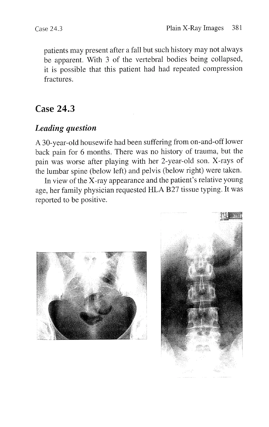
the lumbar spine (below left) and pelvis (below right) were taken.
In view of the X-ray appearance and the patient’s relative young
age, her family physician requested HLA B27 tissue typing. It was
reported to be positive.
View original page (figures / layout)
Page 7
382 Problem-Based Medical Case Management Case 24.3
Questions
1. Describe the most prominent radiological abnormality.
2. What is the radiological diagnosis?
3. What is the underlying diagnosis of this patient’s lower back
pain?
Answers
1. Periarticular osteosclerosis of both sacroiliac joints.
2. Osteitis condensa.
3. Simple mechanical back pain.
Description and discussion
° The anteroposterior view of this patient’s lumbar spine X-ray
appears normal. However, X-ray of the pelvis shows periarticu-
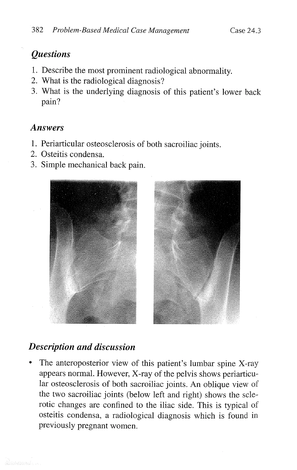
lar osteosclerosis of both sacroiliac joints. An oblique view of
the two sacroiliac joints (below left and right) shows the scle-
rotic changes are confined to the iliac side. This is typical of
osteitis condensa, a radiological diagnosis which is found in
previously pregnant women.
View original page (figures / layout)
Page 8
Case 24.3 Plain X-Ray Images 383
° The underlying pathology of osteitis condensa is poorly under-
stood, but it is often an incidental radiological finding with poor
correlation with the patient’s symptoms.
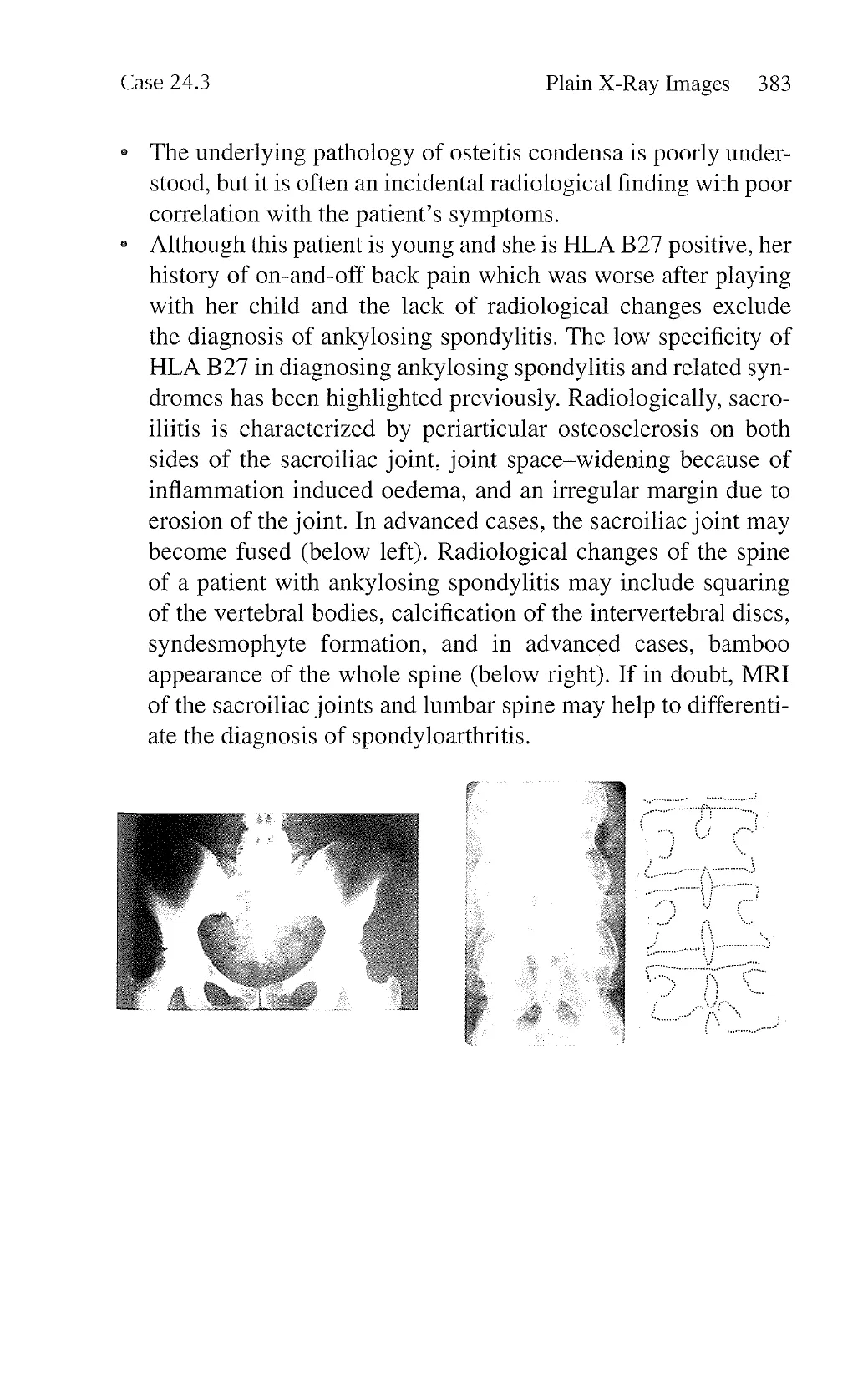
¢ Although this patient is young and she is HLA B27 positive, her
history of on-and-off back pain which was worse after playing
with her child and the lack of radiological changes exclude
the diagnosis of ankylosing spondylitis. The low specificity of
HLA B27 in diagnosing ankylosing spondylitis and related syn-
dromes has been highlighted previously. Radiologically, sacro-
iliitis is characterized by periarticular osteosclerosis on both
sides of the sacroiliac joint, joint space-widening because of
inflammation induced oedema, and an irregular margin due to
erosion of the joint. In advanced cases, the sacroiliac joint may
become fused (below left). Radiological changes of the spine
of a patient with ankylosing spondylitis may include squaring
of the vertebral bodies, calcification of the intervertebral discs,
syndesmophyte formation, and in advanced cases, bamboo
appearance of the whole spine (below right). If in doubt, MRI
of the sacroiliac joints and lumbar spine may help to differenti-
ate the diagnosis of spondyloarthritis.
View original page (figures / layout)
Page 9
384 Problem-Based Medical Case Management Case 24.4
Case 24.4
Leading question
A 55-year-old woman had been complaining of pain and swelling
of multiple joints for 10 years. She had always consulted her local
family physician, who prescribed her with simple analgesics and
non-steroidal anti-inflammatory drugs. The response had always
been suboptimal, but the patient refused referral to a specialist.
Recently, her arthritis had become unbearable. She was admitted
to a local hospital for further management. X-rays of the hands and
feet were taken.
View original page (figures / layout)
Page 10
Case 24.4 Plain X-Ray Images 385
Questions
1.
2.
3.
Describe three radiological abnormalities.
Name two differential diagnoses.
What other physical signs would you look for?
Answers
I.
[SSNS)
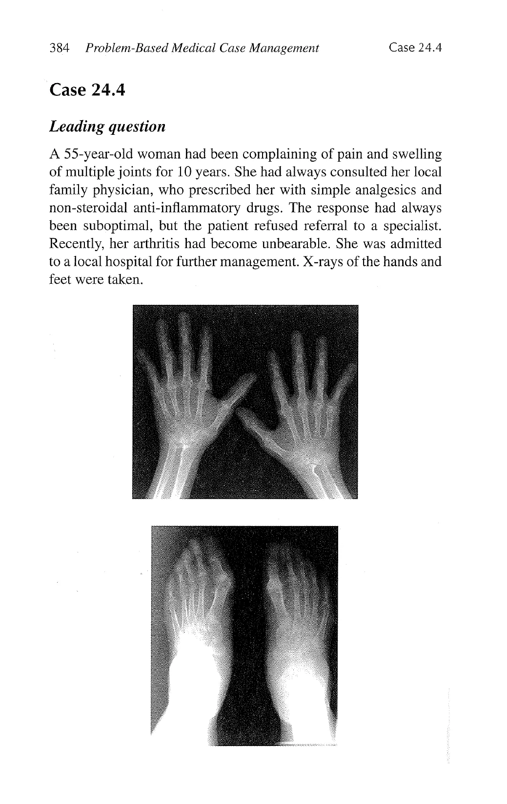
On the hand X-rays, there is periarticular osteopenia; loss of
joint space and periarticular bony erosions involving multiple
joints including the wrist, metacarpophalangeal, and proximal
interphalangeal joints. Corresponding changes are seen on
X-rays of the feet.
. Psoriatic arthritis; rheumatoid arthritis.
. Por psoriatic arthritis: dystrophic nail lesions such as pitting
and hyperkeratosis and scaly erythematosus skin lesions which
are commonly found on the scalp, extensor surfaces of both
upper and lower limbs, anterior surface of both shins, lower
back and peri-umbilical region.
For rheumatoid arthritis: subcutaneous nodules on the ulnar
border of elbows and forearms and other pressure sites; and
other extra-articular manifestations such as digital vasculitis
and pulmonary fibrosis.
Description and discussion
Radiological changes of theumatoid arthritis and psoriatic arthri-
tis may be indistinguishable although bony resorption is more
commonly seen in the latter condition. Additionally, psoriatic
arthritis may sometimes affect the distal interphalangeal joints,
which is uncommon in rheumatoid arthritis.
View original page (figures / layout)
Page 11
386 Problem-Based Medical Case Management Case 24.5
Case 24.5
Leading question
A 65-year-old man consulted his general practitioner because he
had noticed progressive finger joint swelling. Other than occa-
sional niggling pain which usually responded to simple analgesics,
the patient was not troubled by his joint condition. X-rays of the
hands were taken.
Questions
1. Describe four radiological abnormalities.
2. What is the diagnosis?
3. Does this patient require regular arthritis treatment?
View original page (figures / layout)
Page 12
Case 24.6 Plain X-Ray Images 387
Answers
1.
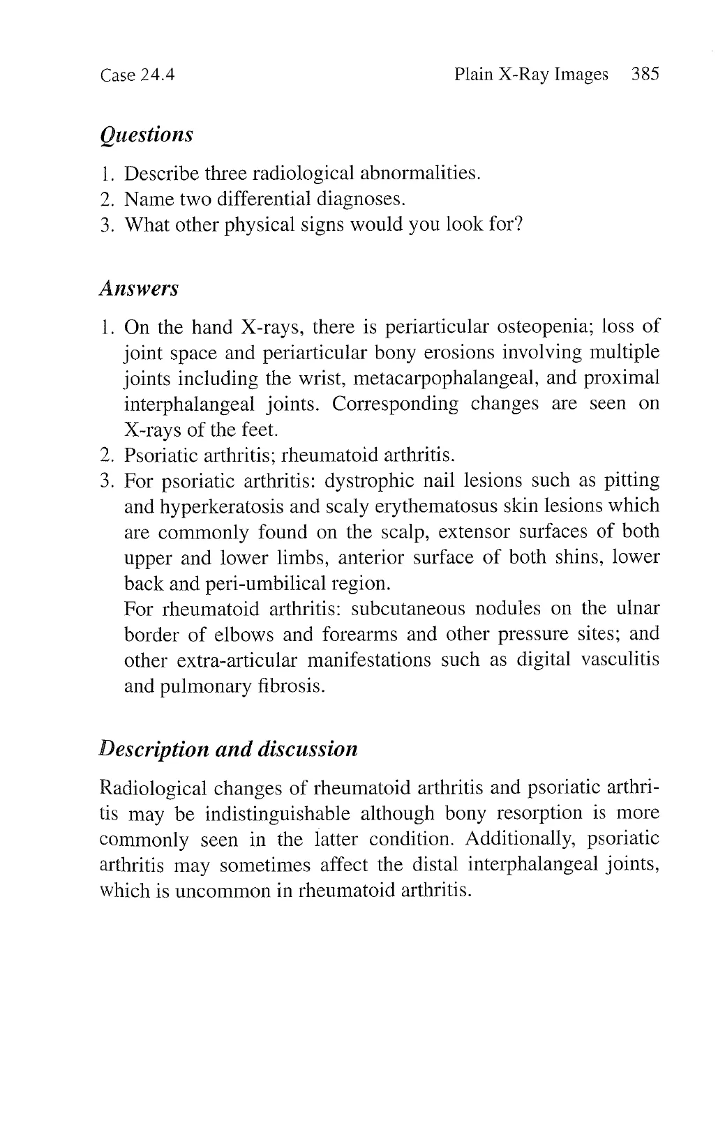
There is loss of joint space, periarticular osteosclerosis, osteo-
phyte formation, and subchondral cyst formation involving
multiple joints including both first carpometacarpal and inter-
phalangeal joints, and the proximal and distal interphalangeal
joints.
. Osteoarthritis.
No.
Description and discussion
This patient’s X-rays exhibit typical changes of osteoarthritis
with loss of joint space, periarticular osteosclerosis, and osteo-
phyte formation. The hand joints that are particularly affected
by osteoarthritis include the first carpometacarpal joint, first
interphalangeal joint, distal interphalangeal joints and, less
commonly, proximal interphalangeal joints. Note that, in con-
trast to rheumatoid arthritis, this patient’s metacarpophalangeal
and wrist joints are spared.
As with osteoarthritis of the other joints, correlation between
radiological changes and the severity of symptoms is poor.
Since this patient is relatively symptom free, there is no need
for any regular medication.
Case 24.6
Leading question
A 45-year-old woman with known rheumatoid arthritis for 10 years
complained of progressive numbness and weakness of all 4 limbs.
A cervical spine X-ray was taken.
View original page (figures / layout)
Page 13
388 Problem-Based Medical Case Management Case 24.6
Questions
1. What view is this cervical spine X-ray?
2. Describe three radiological abnormalities.
3. What other radiological investigation would you recommend?
View original page (figures / layout)
Page 14
Case 24.6 Plain X-Ray Images 389
Answers
1.
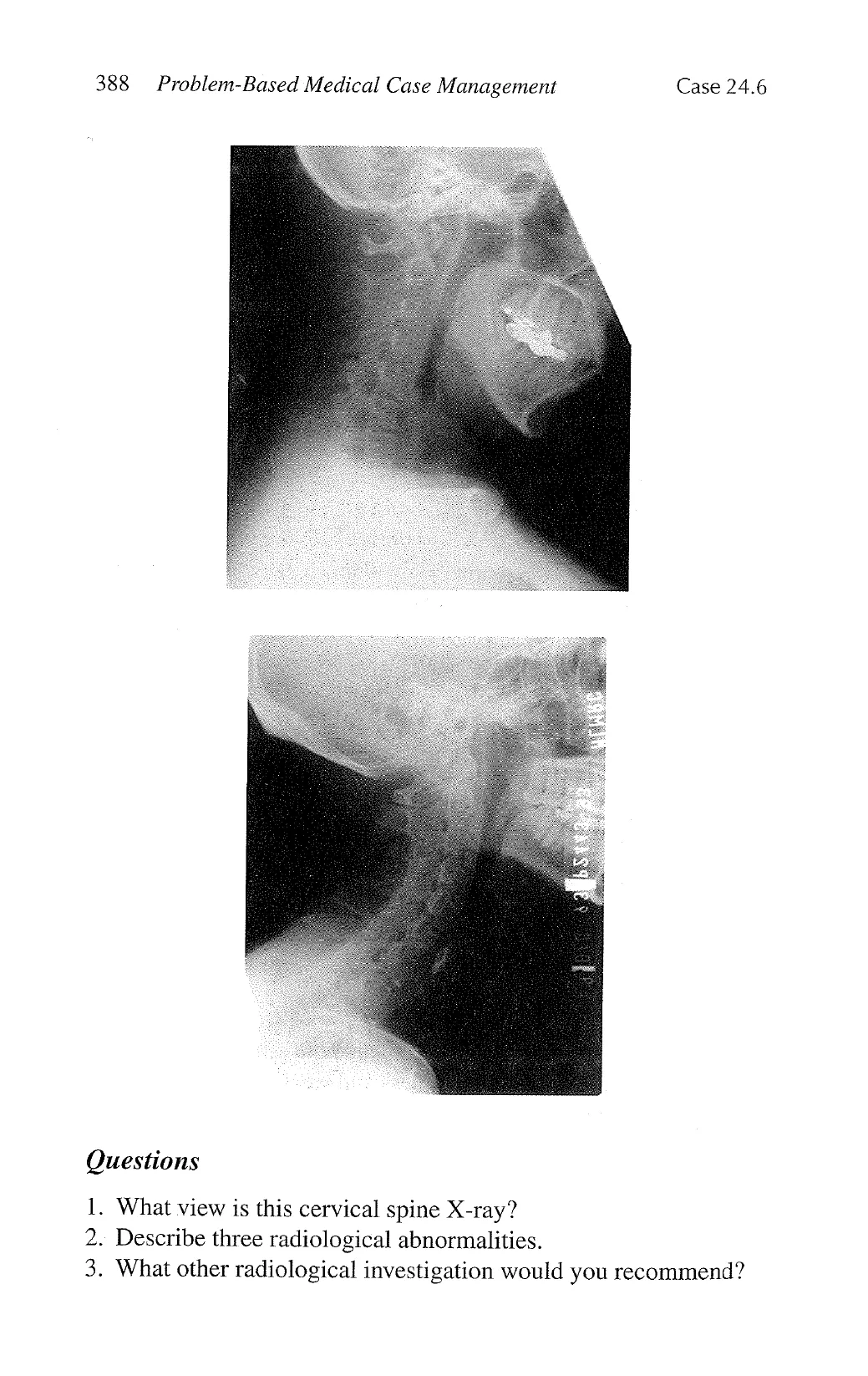
Lateral flexion view.
2. Generalized osteopenia; erosion of the odontoid process axis
and the anterior arch of the first cervical vertebral body (atlas)
and atlantoaxial subluxation.
Magnetic resonance imaging of the cervical spine to look for
impingement of the spinal cord by the odontoid process.
Description and discussion
The clinical differential diagnoses here include cervical
myelopathy and mononeuritis multiplex, both of which are
complications of rheumatoid arthritis. The former is caused by
subluxation of the atlantoaxial joint (C1/2 subluxation) result-
ing in displacement of the odontoid process either upperwardly
or posteriorly compressing on the cervical cord. Mononeuritis
multiplex may be secondary to a vasculitic neuropathy which
is an uncommon complication of rheumatoid arthritis. Patients
may also manifest other extra-articular disease such as rheu-
matoid nodules, vasculitic skin rashes, digital vasculitis, and
pulmonary fibrosis.
To look for possible C1/2 subluxation on a plain X-ray, it is
essential to ask for a lateral flexion view of the cervical spine
although forced flexion should be avoided. A lateral extension
view may not demonstrate the odontoid process displacement.
On this X-ray, there is advanced rheumatoid arthritis involve-
ment of the atlantoaxial joint. The articular surface of the ante-
rior arch of C1 (atlas) appears irregular due to erosions. The
odontoid process of C2 (axis) is poorly visible and is likely to
have been eroded by the rheumatoid process. The axis is dis-
placed posteriorly and may be compressing on the spinal cord.
The odontoid process of the axis may also be displaced
upwardly. Radiologically, this is determined by drawing a line
between the posterior edge of the hard palate and the posterior
edge of the foramen magnum—the chamberlain line. The tip of
the odontoid process should normally be not more than 2.5 mm
above this line.
View original page (figures / layout)
Page 15
390 Problem-Based Medical Case Management Case 24.6
* Plain X-ray appearance of C1/2 subluxation does not confirm
the diagnosis of cervical myelopathy. This should be evaluated
using magnetic resonance imaging, which is capable of visual-
izing the spinal cord and whether there is compression by the
odontoid process.
View original page (figures / layout)