Page 1
22
Skin
Johnny Chun-Yin Chan, Chi-Keung Yeung,
and Henry Hin-Lee Chan
Case 22.1
Leading question
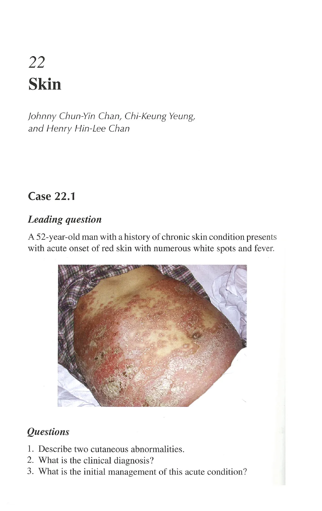
A 52-year-old man with a history of chronic skin condition presents
with acute onset of red skin with numerous white spots and fever.
Questions
1. Describe two cutaneous abnormalities.
2. What is the clinical diagnosis?
3. What is the initial management of this acute condition?
View original page (figures / layout)
Page 2
Case 22.1 Skin 329
Answers
1. Multiple pustules on an erythematous base and multiple well-
demarcated plaques with thick whitish scales on the trunk.
2. Pustular psoriasis.
3. Acutely ill patients with generalized eruption should be hos-
pitalized. Sepsis workup including blood culture and skin
swab, intravenous fluid replacement, correction of electrolyte
disturbance, especially hypocalcaemia, bed rest, and empirical
antibiotics. Topical corticosteroids for the acutely inflamed skin.
Description
¢ Typically presented with sudden onset of burning erythema
that spread rapidly to involve large areas. Tiny pustules then
appear within the bright erythematous areas. The pustules may
coalesce to form bags of pus and the epidermis may slough off,
resulting in erosions.
* Characteristic psoriatic lesions may become inconspicuous
during this acute flare, nail changes may be the only clues for
pre-existing psoriasis.
* Patients often appear very ill, feverish with leukocytosis.
Discussion
* Acute generalized pustular psoriasis is a serious complication
of psoriasis.
* It can be precipitated by withdrawal of systemic corticosteroid
given for the wrong diagnosis of steroid-responding dermatosis.
* Owing to the neutrophilia and fever, sepsis should be excluded
by blood culture and appropriate swabs at the outset.
* Subsequent management should include early referral to der-
matologists for confirmation of diagnosis and commencement
of specific treatment such as systemic retinoids or methotrexate.
Potentially irritating treatments such as tar and phototherapy
should be avoided.
View original page (figures / layout)
Page 3
330 Problem-Based Medical Case Management Case 22.2
* Drug reaction remains an important differential diagnosis of a
generalized pustular eruption.
¢ Erythroderma.
¢ Exfoliative dermatitis.
Case 22.2
Leading question
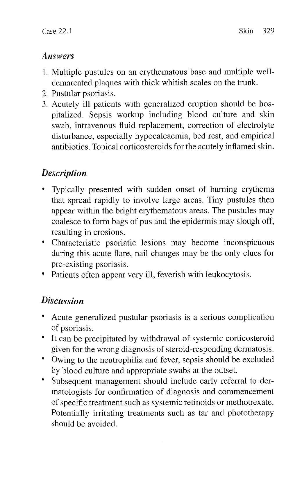
A 38-year-old man with a history of acute myeloid leukaemia
on chemotherapy presents with rapid onset of generalized bright
redness of the skin with chills and rigours.
Questions
1. What is the diagnosis of his skin condition?
2. What are the common causes of this condition?
3. Please outline the initial management of this condition.
View original page (figures / layout)
Page 4
Case 22.2 Skin 331
Answers
1.
. Psoriasis, eczema (atopic dermatitis/contact dermatitis), drug
2
Erythroderma resulted from allergy to systemic antibiotic.
eruption, cutaneous lymphoma.
. Patient should be hospitalized, sepsis workup, correction of
fluid and electrolyte imbalance, constant ambient temperature,
nutritional supplement, emollients, and topical corticosteroids.
Description
Erythroderma is a syndrome of universal erythema of over 95%
of body surface areas. Heavy scaling, dryness, and hyperpig-
mentation are prominent features in chronic stage.
The condition is also known as exfoliative dermatitis.
There may be marked thickening and fissuring of the palms and
soles, nail dystrophy, and thinning of the hair.
It can be associated with systemic upset with fever or hypo-
thermia, regional lymphadenopathy, and dependent oedema.
Hepatosplenomegaly may be found in cutaneous T-cell
lymphoma.
Features of pre-existing dermatosis may only be apparent in the
early stages.
Discussion
.
Erythroderma represents a reaction pattern of the skin and
is potentially life-threatening secondary to failure of skin
functions.
Evaluation of underlying aetiology is essential to control this
serious condition. Peripheral blood smear for atypical lympho-
cytes and skin biopsy are initial investigations.
The most common drugs associated with erythroderma
include antibiotics, especially vancomycin, allopurinol, and
anti-convulsants.
View original page (figures / layout)
Page 5
332 Problem-Based Medical Case Management Case 22.3
e Complications comprise hypoalbuminaemia due to protein loss,
high output cardiac failure, hypothermia, secondary sepsis, par-
ticularly Staphylococcus aureus.
e Systemic absorption of topically applied medications is
enhanced due to large surface area affected and vasodilatation
of capillaries. Appropriate strength of topical steroid should
be used.
Case 22.3
Leading question
A 28-year-old man with a history of allogeneic bone marrow trans-
plantation for lymphoma develops a painful rash on the right flank
for 3 days.
Questions
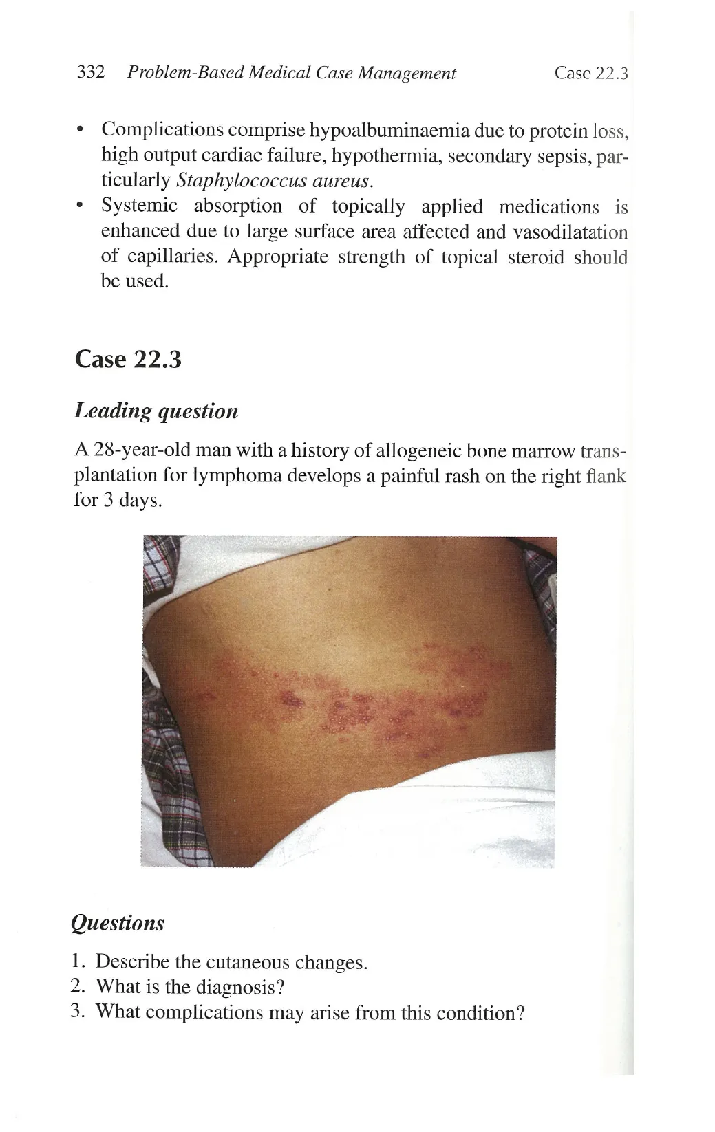
1. Describe the cutaneous changes.
2. What is the diagnosis?
3. What complications may arise from this condition?
View original page (figures / layout)
Page 6
Case 22.3 Skin 333
Answers
1. Multiple vesicles in clusters on erythematous base distributed
along right T10 dermatome.
. Herpes zoster on right T10 dermatome.
. Post-herpetic neuralgia, secondary bacterial infection, and cuta-
neous dissemination.
0o
Description
* Herpes zoster is due to reactivation of varicella-zoster virus
arising from a sensory ganglion. It is characterized by a unilat-
eral vesicular eruption in crops and pain along the correspond-
ing dermatome.
* Preceded by pain or paraesthesia in the involved dermatome
for more than a week before the onset of eruption, mimicking
angina pectoris or acute abdomen.
* Erythematous papules rapidly evolve into vesicles in clusters
and pustules in linear distribution are characteristics of herpes
zoster. In severe cases, haemorrhagic blisters or crusting may
develop, leaving residual dyspigmentation and scarring.
* Common sites involved are thoracic, trigeminal followed by
lumbosacral region.
* Spreading beyond the original dermatome and secondary dis-
semination can occur in immunocompromised patients.
Discussion
* Herpes zoster signifies decrease in specific immunity against
varicella-zoster virus. It is associated with malignancy, espe-
cially lymphoproliferative disorders, and human immuno-
deficiency virus infection, and is more prevalent in organ
transplant recipients and patients on immunosuppressants such
as corticosteroids.
° Search for the cause of depressed immunity is necessary espe-
cially in patients younger than 55 years of age.
View original page (figures / layout)
Page 7
334 Problem-Based Medical Case Management Case 22.4
e Oral or systemic antiviral therapy, such as acyclovir, can be
given depending on the background immunocompromised
state. It also helps to reduce duration of eruption and post-
herpetic neuralgia if given in early stage.
e Topical or oral antibiotics can be used to treat secondary bacte-
rial infection and adequate analgesic should be given.
¢ Ophthalmologists should be consulted for ophthalmic zoster for
the risk of keratitis and other ocular complications.
e Vasculitis.
Case 22.4
Leading question
A 51-year-old woman presents with rash on both legs associated
with ankle joint pain and swelling.
Questions
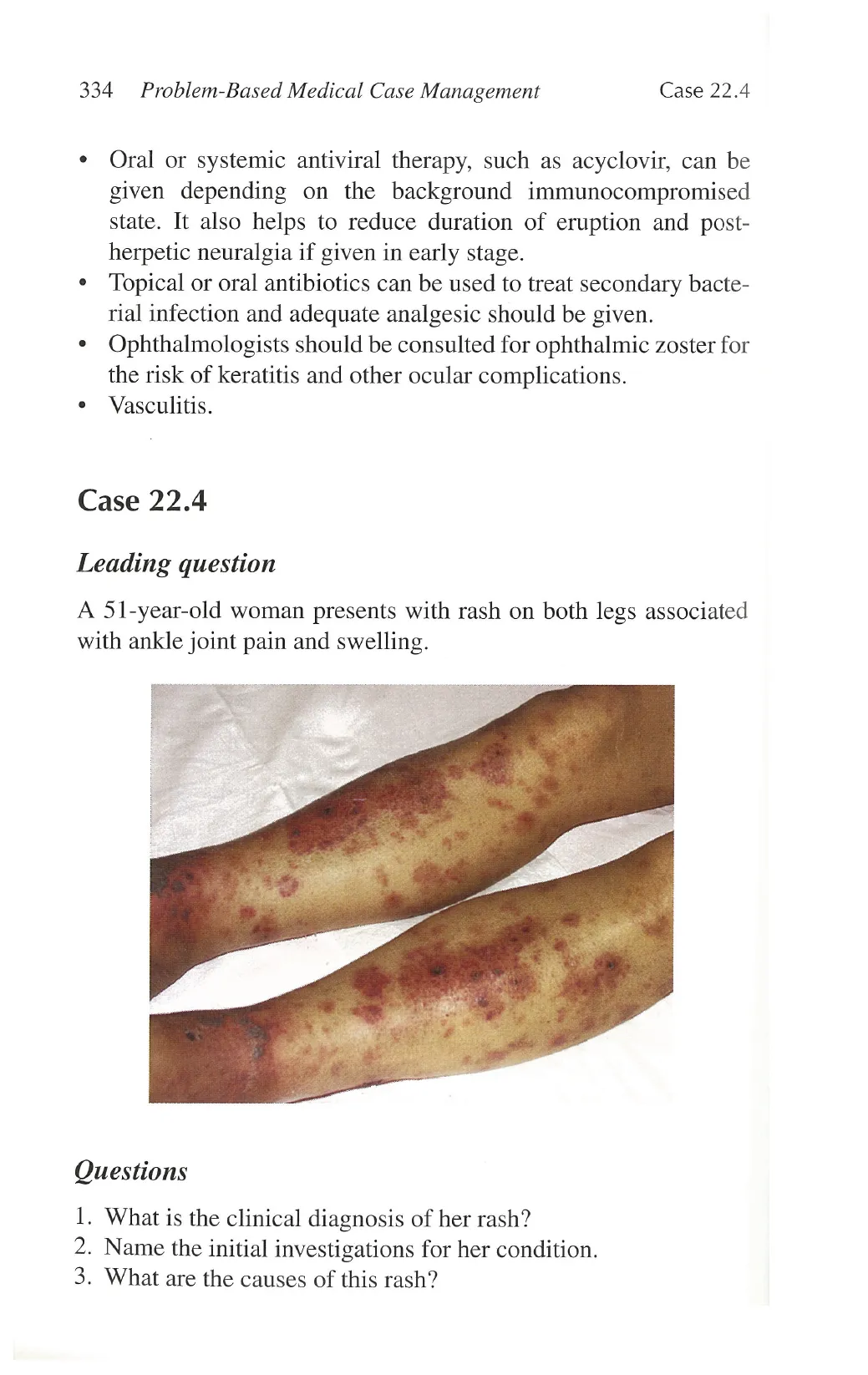
1. What is the clinical diagnosis of her rash?
2. Name the initial investigations for her condition.
3. What are the causes of this rash?
View original page (figures / layout)
Page 8
Case 22.4 Skin 335
Answers
1.
S
The clinical diagnosis of multiple palpable purpura is leukocy-
toclastic vasculitis or allergic angiitis.
Complete blood counts, liver and renal function tests, ESR,
clotting profiles, vasculitis markers, such as ANA, RF, ANCA,
C3, C4, immunoglobulin pattern, skin biopsy with direct
immunoflorescence, urinanalysis.
. Henoch-Schonlein purpura occurs mostly in children, viral
infections, bacterial infections such as Group A streptococ-
cus, tuberculosis, drug reaction, systemic lupus, haemato-
logical malignancy, rheumatoid arthritis, cryoglobulinaemia,
paraproteinaemia.
Description
Typical features are palpable non-blanchable purpura on
dependent areas, mainly lower legs and ankles. Florid lesions
can develop into blisters and erosions.
Distinct from the flat purpura due to platelet and clotting
abnormalities.
Associated with polyarthralgia and ankle swelling.
The associated features depend on the underlying aetiology,
history of recent drug intake, symptoms of upper respiratory
tract infection, underlying connective tissue disorders.
In acutely ill patients, bacteraemia, septic vasculitis and dis-
seminated intravascular coagulation must be ruled out.
Discussion
There are diverse causes of vasculitis associated with hyper-
sensitivity to various antigens. The presenting features depend
on size of involved blood vessels. Inflammation and fibrinoid
necrosis of postcapillary venules are found in leukocytoclastic
vasculitis.
Screening for internal organ involvement is essential, particu-
larly kidneys, manifesting as abnormal urine sediments, azotae-
mia and hypertension, gut, joint and peripheral nerves.
View original page (figures / layout)
Page 9
336 Problem-Based Medical Case Management Case 22.5
* Treatment is based on underlying cause: antibiotics for bacte-
rial causes, systemic corticosteroid, and other immunosuppres-
sants may be indicated for internal organ involvement. Second
line therapy includes dapsone and colchicine.
e Cellulitis.
Case 22.5
Leading question
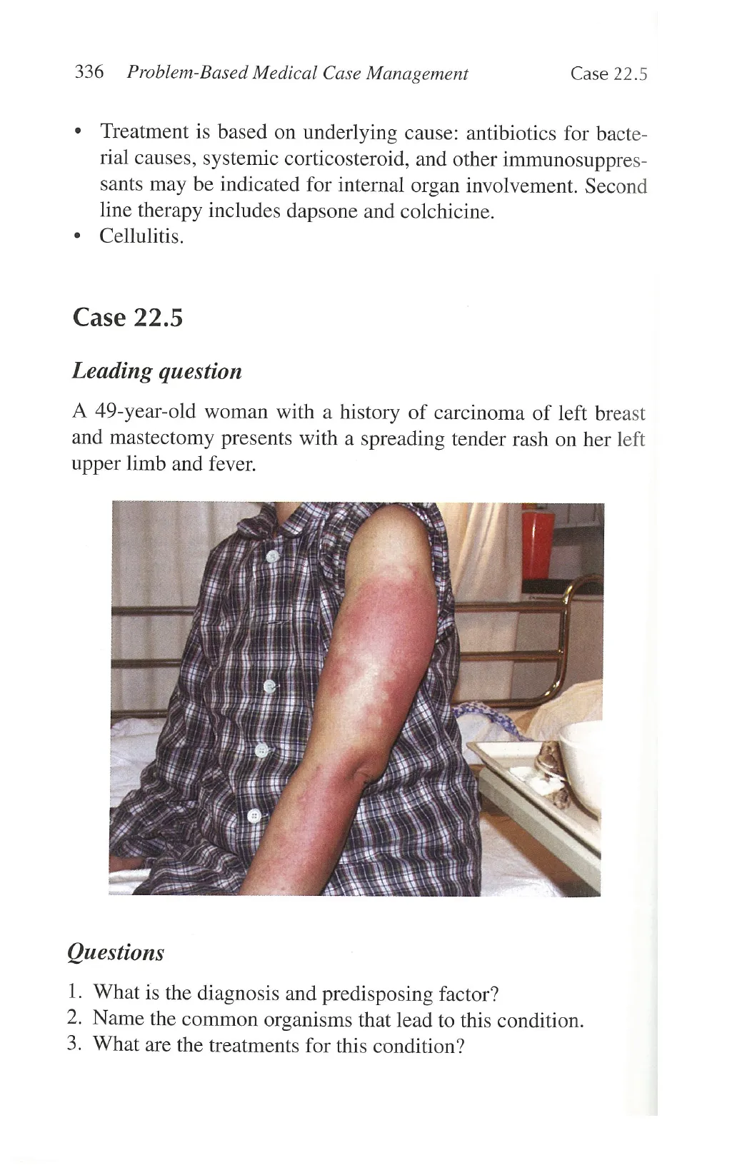
A 49-year-old woman with a history of carcinoma of left breast
and mastectomy presents with a spreading tender rash on her left
upper limb and fever.
Questions
1. What is the diagnosis and predisposing factor?
2. Name the common organisms that lead to this condition.
3. What are the treatments for this condition?
View original page (figures / layout)
Page 10
Case 22.5 Skin 337
Answers
1.
o
Cellulitis of the left upper limbs resulted from secondary lym-
phoedema after mastectomy.
. Group A B-haemolytic Streptococci, Staphylococcus aureus
and Haemophilus influenzae in children.
. Intravenous ampicillin and cloxacillin for 1-2 weeks and surgi-
cal drainage if abscesses develop. Erythromycin can be used in
patients with penicillin allergy.
Description
Cellulitis usually presents with classical signs of acute inflam-
mation, namely, redness, tenderness, warmth, and swelling.
Blisters, lymphangitis with erythematous streaking and regional
Iymphadenopathy may be present.
In moderate to severe cellulitis, patients often develop fever and
systemic upset.
Predisposing factors may be obvious, such as tinea paedis,
eczema, blisters due to herpes infections, lymphoedema sec-
ondary to operation and radiotherapy, ischemic ulcers, trauma,
surgical wound, intravenous drug users, and diabetes mellitus.
Most common sites are the legs and face.
Cellulitis can be complicated by necrotizing fasciitis that
carries high mortality, and should not be overlooked..Patients
with necrotizing fasciitis often appear very ill with high fever
and unstable haemodynamic status; the degree of pain can be
disproportional to the apparent cutaneous changes.
Discussion
Cellulitis is acute spreading inflammation of dermis and subcu-
taneous tissues due to bacterial invasion.
Differential diagnosis is deep vein thrombosis if lower limbs
are affected.
View original page (figures / layout)
Page 11
338 Problem-Based Medical Case Management Case 22.6
e Underlying predisposing factors should be sought to prevent
recurrence of cellulitis that would lead to further destruction of
lymphatics and chronic lymphoedema.
¢ Investigation includes wound swab for culture, blood culture,
and white cell count. X-ray may be needed for assessing under-
lying soft tissue and bone involvement.
Case 22.6
Leading question
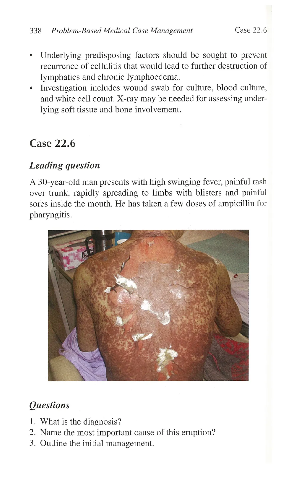
A 30-year-old man presents with high swinging fever, painful rash
over trunk, rapidly spreading to limbs with blisters and painful
sores inside the mouth. He has taken a few doses of ampicillin for
pharyngitis.
Questions
1. What is the diagnosis?
2. Name the most important cause of this eruption?
3. Outline the initial management.
View original page (figures / layout)
Page 12
Case 22.6 Skin 339
Answers
1. Toxic epidermal necrolysis.
2. Drug hypersensitivity. Ampicillin is the culprit drug in this
case.
3. Early withdrawal of the potential responsible medication is the
most important step. The patient is to be admitted to a burn
unit and treated as a second-degree burn patient. Regular sepsis
surveillance and appropriate antibiotics, correction of fluid and
electrolyte imbalance and nutritional supplement are important.
Description
+ Toxic epidermal necrolysis presents initially with flu-like symp-
toms such as fever and sore throat. Patients then develop painful
lip erosions and swelling with sore mouth and eyes, followed
by spreading tender erythema of the trunk and face. The skin
soon becomes necrotic and develops blisters with extensive
erosions.
* Conjunctiva and oral mucosa are most commonly affected with
painful erosions.
* Stevens-Johnson syndrome differs from toxic epidermal
necrolysis only by a lesser extent of body surface area involved
(< 10%). Erythema multiforme is now considered to be a differ-
ent entity. It typically manifests as target lesions mostly on the
acral areas in contrast to truncal involvement in toxic epidermal
necrolysis.
Discussion
* Toxic epidermal necrolysis is the most severe mucocutaneous
reaction to drug with significant mortality from multisystem
involvement and sepsis.
Most frequently related drugs are sulphonamides, allopurinol,
anticonvulsants, non-steroidal anti-inflammatory drugs, and
penicillins.
View original page (figures / layout)
Page 13
340 Problem-Based Medical Case Management Case 22.7
e Staphylococcal scalded skin syndrome is the main differential
diagnosis, but it is mostly found in children and immunocom-
promised patients.
e There is no universally accepted effective treatment for toxic
epidermal necrolysis. Systemic corticosteroid is given only in
selected cases because of its side effects. Intravenous immu-
noglobulin seems to be a promising therapy in halting the
blistering process and reducing the mortality, based on several
reported series.
¢ Alert to avoid offending drugs to prevent fatal outcome of read-
ministering the medication.
e Panniculitis.
¢ Erythema nodosum.
Case 22.7
Leading question
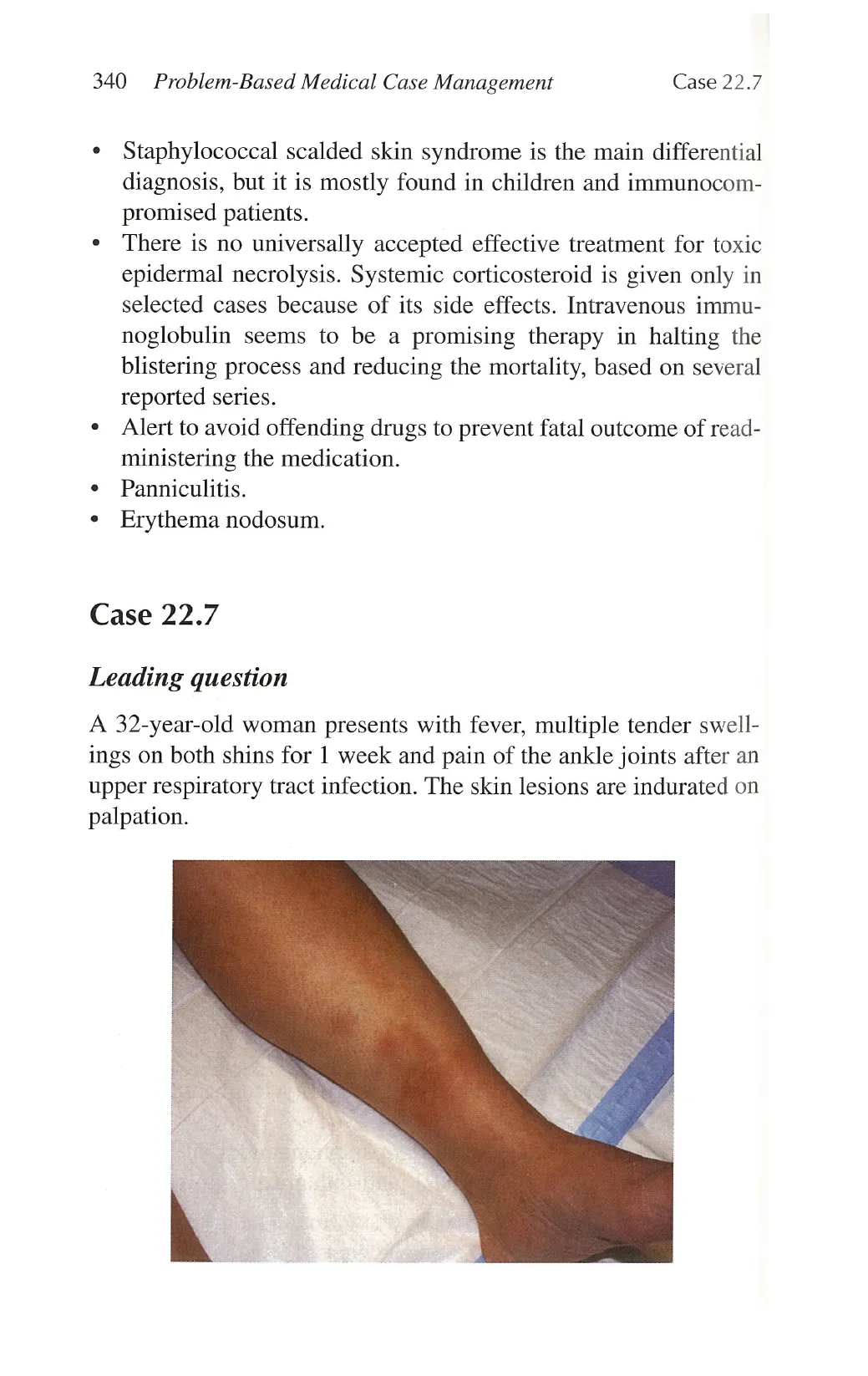
A 32-year-old woman presents with fever, multiple tender swell-
ings on both shins for 1 week and pain of the ankle joints after an
upper respiratory tract infection. The skin lesions are indurated on
palpation.
View original page (figures / layout)
Page 14
Case 22.7 Skin 341
Questions
1. What is the clinical diagnosis?
2. Name five common associations with this condition.
3. Outline the management of this condition.
Answers
1. Erythema nodosum or panniculitis.
2. Tuberculosis, drugs, Streptococcal infection, inflammatory
bowel disease, and Bechet’s syndrome.
3. Treat the underling cause, bed rest and non-steroidal anti-
inflammatory drugs.
Description
* Multiple discrete indurated tender erythematous nodules on
both legs, especially in the pretibial region, and associated with
painful swelling of the ankles. The lesions may also appear on
knees and arms.
* The nodules usually resolve with bruise-like post-inflammatory
hyperpigmentation within weeks.
* Differential diagnoses include other types of panniculitis,
medium-sized vasculitis, and thrombophlebitis.
* Idiopathic cases are more prevalent in young women.
Discussion
* Erythema nodosum is an acute inflammation of the subcutane-
ous fat, predominantly involving the septum without vasculitis.
* Thisis areaction pattern to a wide range of stimuli. Leukocytosis
and raised inflammatory markers are evident by blood tests.
* It is important to look for tuberculosis in the high prevalence
areas. Chest signs, regional lymphadenopathy, and constitu-
tional symptoms are to be sought. Investigations should include
chest X-ray and Mantoux test.
View original page (figures / layout)
Page 15
342 Problem-Based Medical Case Management Case 22.8
¢ Oral contraceptives and sulphonamides are frequently responsi-
ble drugs.
« Skin biopsy is indicated for recurrent cases to differentiate from
other types of panniculitis and vasculitis.
¢ Pemphigus vulgaris.
Case 22.8
Leading question
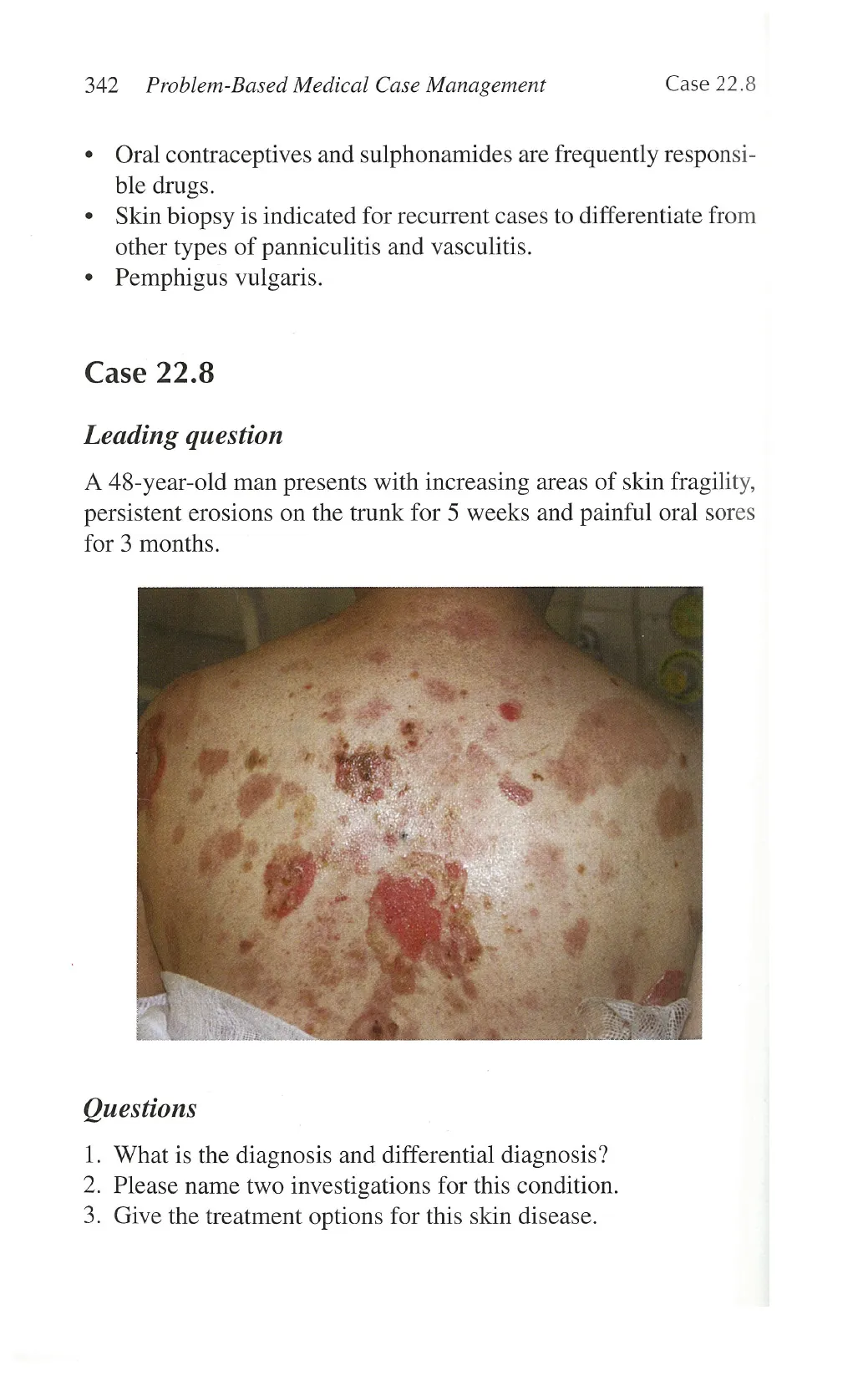
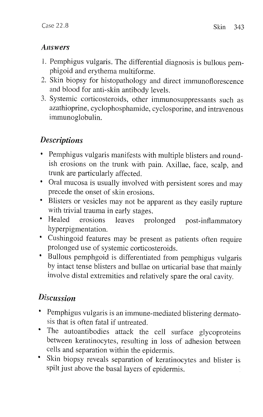
A 48-year-old man presents with increasing areas of skin fragility,
persistent erosions on the trunk for 5 weeks and painful oral sores
for 3 months.
Questions
1. What is the diagnosis and differential diagnosis?
2. Please name two investigations for this condition.
3. Give the treatment options for this skin disease.
View original page (figures / layout)
Page 16
Case 22.8 Skin 343
Answers
o
Pemphigus vulgaris. The differential diagnosis is bullous pem-
phigoid and erythema multiforme.
. Skin biopsy for histopathology and direct immunoflorescence
and blood for anti-skin antibody levels.
. Systemic corticosteroids, other immunosuppressants such as
azathioprine, cyclophosphamide, cyclosporine, and intravenous
immunoglobulin.
Descriptions
Pemphigus vulgaris manifests with multiple blisters and round-
ish erosions on the trunk with pain. Axillae, face, scalp, and
trunk are particularly affected.
Oral mucosa is usually involved with persistent sores and may
precede the onset of skin erosions.
Blisters or vesicles may not be apparent as they easily rupture
with trivial trauma in early stages.
Healed erosions leaves prolonged post-inflammatory
hyperpigmentation.
Cushingoid features may be present as patients often require
prolonged use of systemic corticosteroids.
Bullous pemphgoid is differentiated from pemphigus vulgaris
by intact tense blisters and bullae on urticarial base that mainly
involve distal extremities and relatively spare the oral cavity.
Discussion
Pemphigus vulgaris is an immune-mediated blistering dermato-
sis that is often fatal if untreated.
The autoantibodies attack the cell surface glycoproteins
between keratinocytes, resulting in loss of adhesion between
cells and separation within the epidermis.
Skin biopsy reveals separation of keratinocytes and blister is
spilt just above the basal layers of epidermis.
View original page (figures / layout)
Page 17
344 Problem-Based Medical Case Management Case 22.9
* Positive immune deposits are found in the intercellular sub-
stance pattern of the epidermis. Autoantibody titers in serum
usually correspond with disease activity.
¢ Systemic immunosuppressants are essential to treat the disease
which is progressive. Prolonged systemic corticosteroid is
the main stay of treatment. Concomitant immunosuppressant
therapy is usually required for steroid-sparing effect.
Case 22.9
Leading Question
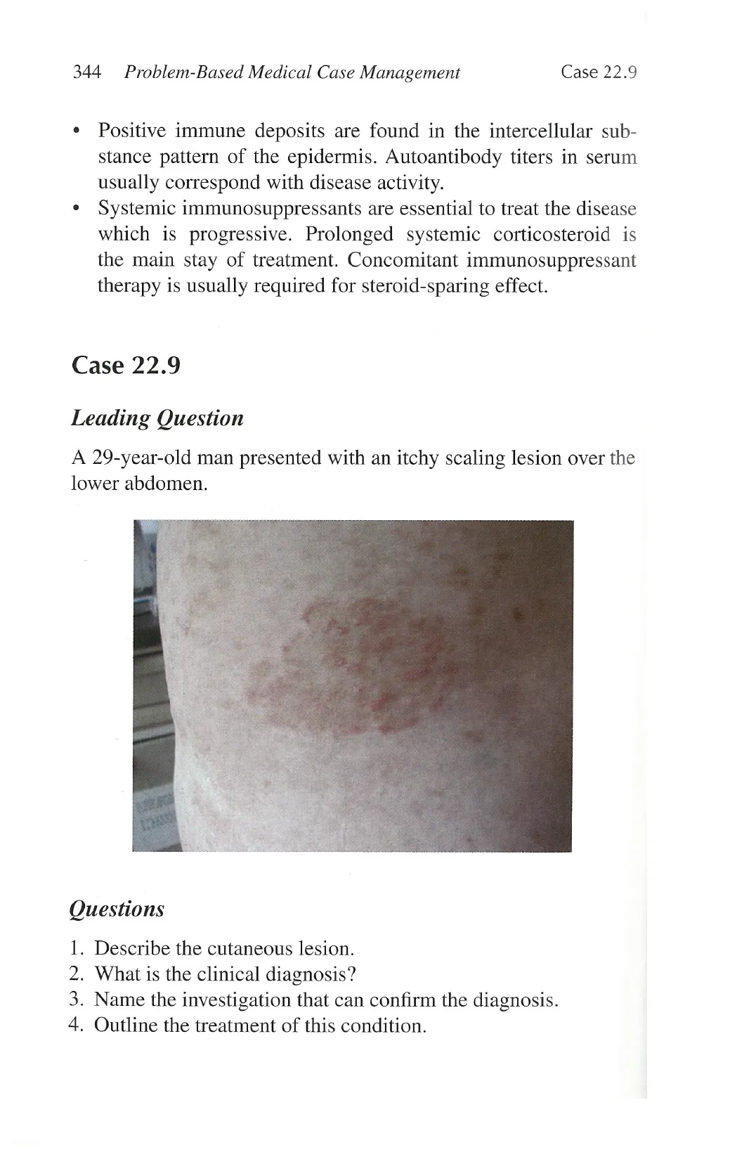
A 29-year-old man presented with an itchy scaling lesion over the
lower abdomen.
Questions
1. Describe the cutaneous lesion.
2. What is the clinical diagnosis?
3. Name the investigation that can confirm the diagnosis.
4. Outline the treatment of this condition.
View original page (figures / layout)
Page 18
Case 22.9 Skin 345
Answers
1.
]
Well-defined, annular, erythematous patch with a scaling border
over the lower abdomen.
Tinea corporis.
Skin scraping for fungus. Superficial scraps of the skin (pre-
pared with potassium hydroxide) showing fungal hyphae under
microscopy will confirm the diagnosis.
Topical anti-fungal agents including the azoles (e.g., clotrima-
zole, tioconazole, isconazole) and allylamines (e.g., terbinafine)
can be used to treat tinea corpotis.
Discussions
1.
Tinea refers to superficial fungal infection. Different names
are used to denote superficial fungal infection at various body
sites (e.g., tinea corporis—glabrous skin over trunk, arms, and
legs; tinea manuum—hands; tinea pedis—soles; tinea cruris—
groin; tinea faciale—face; tinea capitis—scalp; tinea unguium
[onychomycosis]—nails).
Tinea is caused by dermatophytes. Dermatophytic fungi include
Trichophyton, Epidermophyton, and Microsporum species.
These fungi infect the most superficial part of the epidermis,
namely, the stratum corneum and nail plates.
. A thorough dermatological examination should be carried out
when a diagnosis of tinea has been made. Involvement of other
body sites is frequent and the reservoir of dermatophytes should
be carefully sought out. Tinea pedis and tinea unguium (onych-
omycosis) are the common reservoirs for recurrent infections.
Systemic anti-fungal agents can be used to treat refractory tinea
not responding to topical therapies. Azoles (e.g., fluconazole,
itraconazole, ketoconazole) and allylamines (e.g., terbinafine)
are effective systemic therapies in eradicating tinea.
View original page (figures / layout)
Page 19
346 Problem-Based Medical Case Management Case 22.10
Case 22.10
Leading question
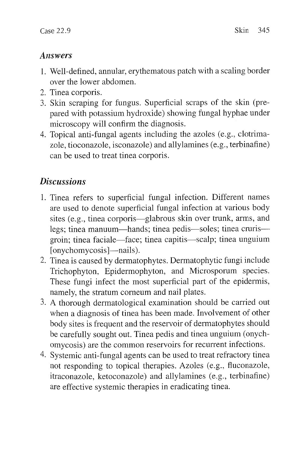
A 24-year-old man presented with a subacute onset of hair loss
over the vertex.
Questions
1. Describe the scalp abnormality.
2. What is the clinical diagnosis?
3. What are the associated cutaneous features?
4. What are the treatment options for the condition?
Answers
1. Multiple well-defined patches of non-scarring alopecia.
2. Alopecia areata.
3. Alopecia areata is associated with nail pitting and increased
incidence of atopic dermatitis.
View original page (figures / layout)
Page 20
Case 22.11 Skin 347
4. Small lesions can be observed and may later regress spontane-
ously with time. Potent topical corticosteroid (e.g., clobetasone
propionate) and topical calcineurin inhibitor (tacrolimus, pime-
crolimus) can be used. Intralesional injection of corticosteroid
(triamcinolone acetonide) is sometimes employed to treat
extensive alopecia areata.
Discussions
1. Alopecia areata is a form of non-scarring alopecia with an auto-
immune origin. It often presents as subacute onset of circular or
oval-shaped patch of hair loss.
2. In severe cases, there can be involvement of the whole scalp
(alopecia totalis) or even the whole body (alopecia universalis).
3. Alopecia areata is epidemiologically linked to other autoim-
mune diseases (including thyroid diseases, systemic lupus ery-
thematosus, rheumatoid arthritis, etc.) and atopic dermatitis.
4. Minoxidil is another useful topical agent in treating alopecia
areata. Systemic immunosuppressants such as cyclosporine
can be used to retard the disease progress in severe diseases.
In patients with extensive alopecia, immunotherapy with di-
phenyl-cyclo-propenone (DPCP) can be an effective therapy by
inducing a local irritant dermatitis reaction over the scalp.
Case 22.11
Leading Question
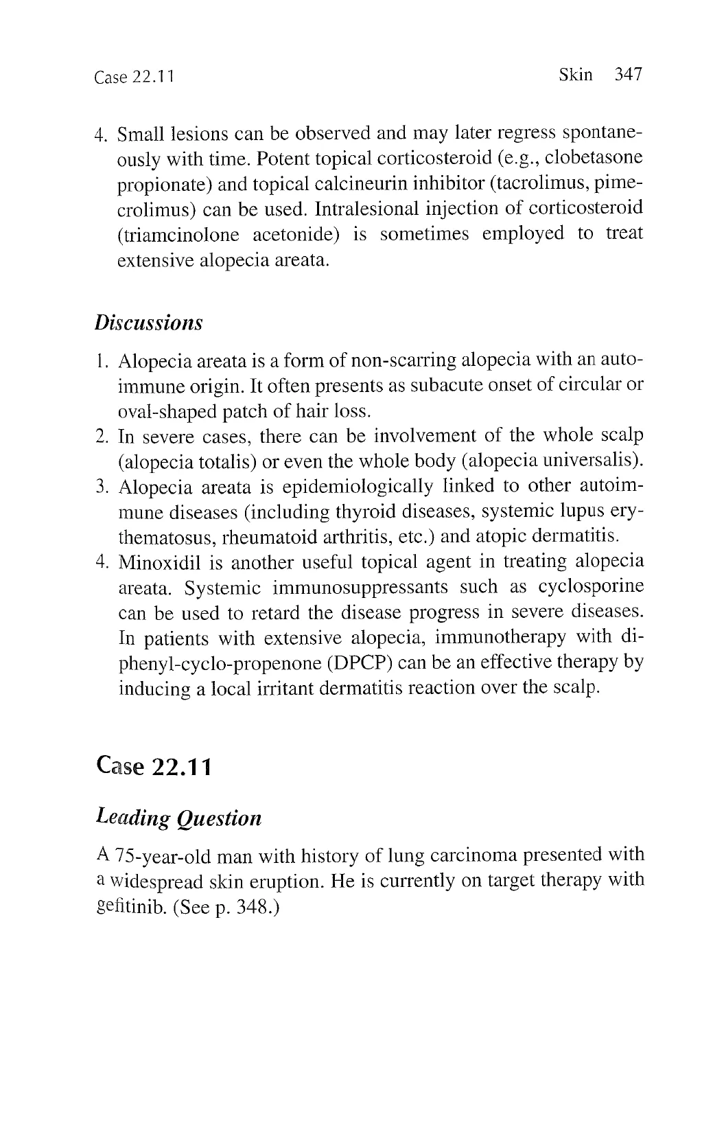
A 75-year-old man with history of lung carcinoma presented with
a widespread skin eruption. He is currently on target therapy with
gefitinib. (See p. 348.)
View original page (figures / layout)
Page 21
348 Problem-Based Medical Case Management Case 22.11
Questions
1. Describe the cutaneous abnormality.
2. What is the clinical diagnosis?
3. Outline the treatment principles of the condition.
Answers
1. Generalized papulopustules over the trunk.
2. Acneiform eruption due to gefitinib.
3. Topical agents including clindamycin, erythromycin, benzoyl
peroxide, and retinoid can be used. Systemic therapies includ-
ing courses of tetracyclines or macrolides will be prescribed in
refractory cases.
Discussions
1. Gefitinib belongs to the group of tyrosine kinase inhibitors
(TKI). Cutaneous adverse effects of TKI include acneiform
View original page (figures / layout)
Page 22
Case 22.12 Skin 349
eruption, scalp dermatitis, xerosis (dryness of skin), asteatotic
eczema, and hand-foot syndrome.
2. The incidence of cutaneous adverse effects of TKI is rising with
increased use of TKI in oncological patients.
3. Tetracyclines and macrolides have intrinsic immunomodula-
tory effects that can treat the acneiform eruption due to TKI.
4. Liberal use of emollients is essential in patients with xerosis and
asteatotic eczema. Potent topical corticosteroid is sometimes
required to treat severe scalp dermatitis and asteatotic eczema.
Case 22.12
Leading Question
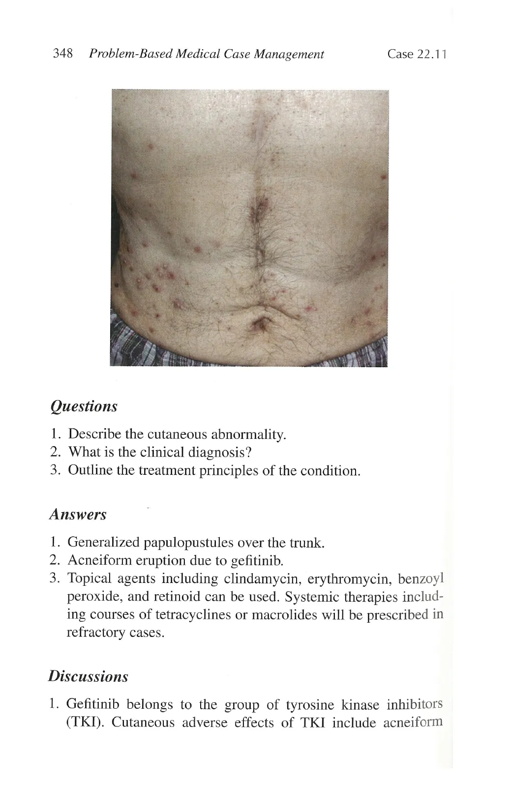
A 57-year-old woman presented with an acute onset of itchy rash
localized over the face. She volunteered history of using a new
facial cleanser.
View original page (figures / layout)
Page 23
350 Problem-Based Medical Case Management Case 22.12
Questions
1.
2.
3.
4.
Describe the cutaneous abnormality on face.
‘What is the clinical diagnosis?
‘What are the important differential diagnoses?
Outline the treatment principles.
Answers
1.
2.
. Differential diagnoses including systemic lupus erythematosus,
There is diffuse erythema all over the face. Scaling is prominent
over the central face.
Contact allergic dermatitis due to the new cleanser.
dermatomyositits, atopic dermatitis, seborrhoeic dermatitis,
rosacea, and angioedema.
. Patients should be advised to avoid future use of the cleanser.
A short course (in days) of topical corticosteroid can be used to
treatment the dermatitis.
Discussions
1.
Contact dermatitis can be classified into allergic type and irri-
tant type. Contact allergic dermatitis is common on the face due
to allergic reactions to products such as cosmetic, cleansers,
masks, sunscreen or eye drops etc.
. Patients will present with erythema, increased warmth, and
swelling over the localized areas in contact with the culprit
agents. Epidermal change (scaling) is an important sign in diag-
nosis contact allergic dermatitis.
. Potent topical corticosteroid is used to treat contact allergic
dermatitis. Caution should be taken as prolonged use of potent
topical corticosteroid could cause skin atrophy on face. -
Skin patch test can be used to identify the culprit of contact
allergic dermatitis. A selected panel of allergens is placed onto” -
the back of the patients. Specific skin reactions will be looked
for at both 48 hours and 96 hours to determine the possible of -
allergy. .
View original page (figures / layout)
Page 24
Case 22.13 Skin 351
5. Skin patch test will give circumstantial evidence of possible
sources of the allergic reaction. Clinical relevance should be
confirmed by the physician in discussing the possible exposure
with the patient.
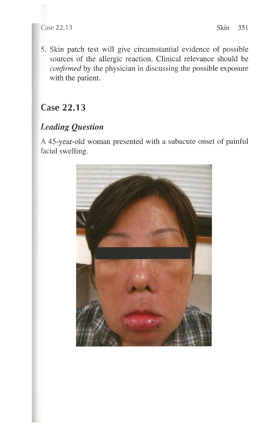
Case 22.13
Leading Question
A 45-year-old woman presented with a subacute onset of painful
facial swelling.
View original page (figures / layout)
Page 25
352 Problem-Based Medical Case Management Case 22.13
Questions
1.
2.
3.
4.
Describe the cutaneous abnormality.
What is the clinical diagnosis?
What are the differential diagnoses?
Outline the treatment principle in this condition.
Answers
1.
2.
3.
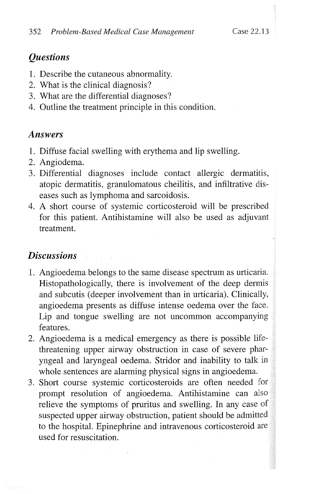
Diffuse facial swelling with erythema and lip swelling.
Angiodema.
Differential diagnoses include contact allergic dermatitis,
atopic dermatitis, granulomatous cheilitis, and infiltrative dis-
eases such as lymphoma and sarcoidosis.
. A short course of systemic corticosteroid will be prescribed
for this patient. Antihistamine will also be used as adjuvant
treatment.
Discussions
1.
Angioedema belongs to the same disease spectrum as urticaria.
Histopathologically, there is involvement of the deep dermis
and subcutis (deeper involvement than in urticaria). Clinically,
angioedema presents as diffuse intense oedema over the face.
Lip and tongue swelling are not uncommon accompanying
features.
. Angioedema is a medical emergency as there is possible life-
threatening upper airway obstruction in case of severe phar-
yngeal and laryngeal oedema. Stridor and inability to talk in
whole sentences are alarming physical signs in angioedema.
. Short course systemic corticosteroids are often needed for
prompt resolution of angioedema. Antihistamine can also
relieve the symptoms of pruritus and swelling. In any case of
suspected upper airway obstruction, patient should be admitted
to the hospital. Epinephrine and intravenous corticosteroid are
used for resuscitation.
View original page (figures / layout)