Page 1
21
Fundi
Nelson Ming-Sun Wat
Case 21.1
Leading question
A 52-year-old man complains of severely impaired visual acuity.
Question
0
What is the diagnosis?
View original page (figures / layout)
Page 2
318 Problem-Based Medical Case Management Case 21.2
Answer
* Atrophic papilloedema.
Description
¢ Very pale white disc, slightly elevated with indistinct margins,
also called “secondary optic atrophy” due to long standing
papilloedema.
Case 21.2
Leading question
A 78-year-old man with a history of atrial fibrillation complains of
sudden visual field loss.
Question
¢ What is the diagnosis?
View original page (figures / layout)
Page 3
Case 21.3 Fundi 319
Answer
Retinal artery occlusion.
Discussion
* What are the causes?
— Emboli.
— Atherosclerosis.
Case 21.3
Leading question
This 67-year-old patient complains of gradual loss of visual acuity.
Question
* What is the diagnosis?
View original page (figures / layout)
Page 4
320 Problem-Based Medical Case Management Case 21.4
Answer
¢ Central retinal vein occlusion.
Description
* There are tortuosity and dilatation of all branches of central
retinal vein, with retinal haemorrhages throughout all 4 quad-
rants. There is optic disc oedema and macular oedema.
Case 21.4
Leading question
A 60-year-old man with a history of diabetes complains of blurring
of vision.
Question
* What is the diagnosis?
View original page (figures / layout)
Page 5
Case 21.5 Fundi 321
Answer
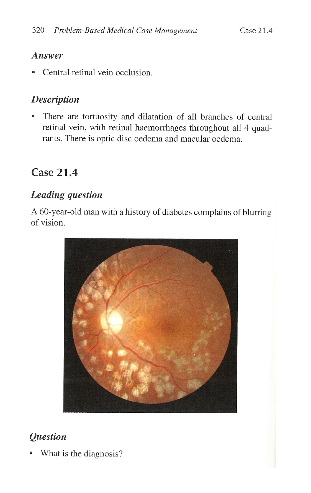
+ Laser photocoagulation scars.
Discussion
¢ Photocoagulation if administered early can prevent blindness
secondary to proliferative diabetic retinopathy. The best results
in proliferative retinopathy are obtained before the onset of
visual symptoms and tractional complications, and in macu-
lopathy before severe visual loss.
Case 21.5
Leading question
A 64-year-old man with a history of hypertension complains of
headache.
Question
What is the underlying condition?
View original page (figures / layout)
Page 6
322 Problem-Based Medical Case Management Case 21.6
Answer
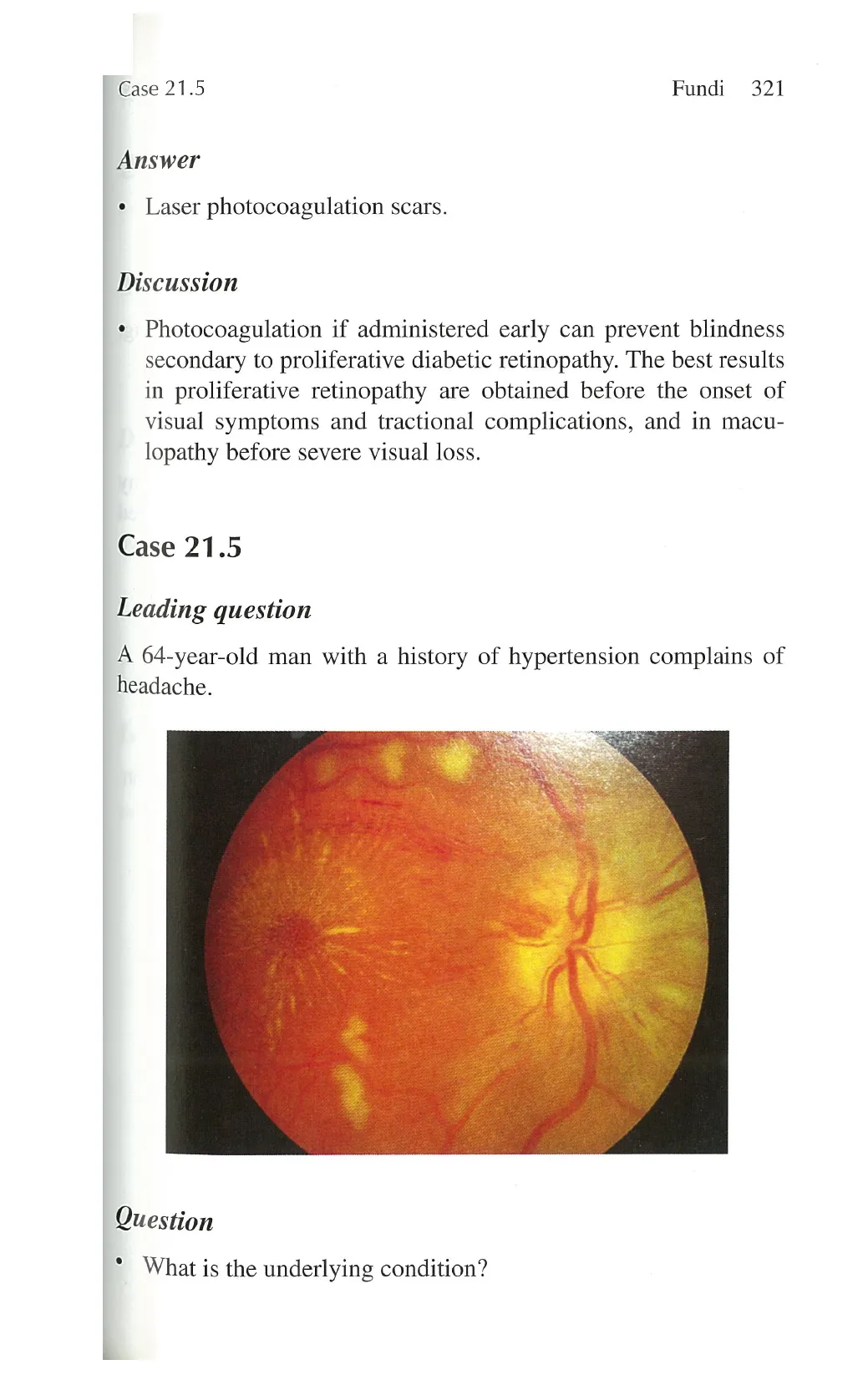
e Malignant hypertension.
Description
¢ Cotton wool spot, flame-shaped haemorrhages, disc swelling,
and macular star.
Discussion
¢ Hypertensive fundi is distinguished from diabetic retinopathy
which has blot and dot haemorrhages and well-circumscribed
hard exudates. Laser scars are often present. However, the two
conditions commonly coexist.
Case 21.6
Leading question
A 65-year-old woman presents with progressive blurring of vision.
View original page (figures / layout)
Page 7
Case 21.7 Fundi 323
Question
» What is your diagnosis?
Answer
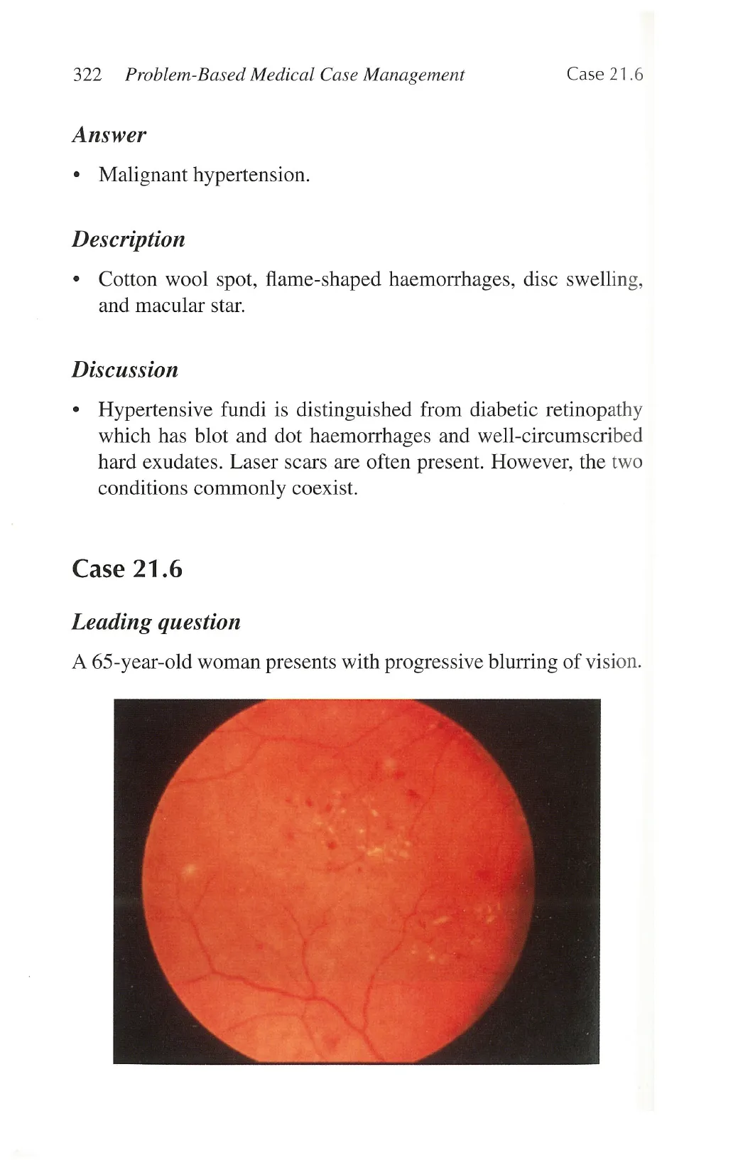
+ Diabetic non-proliferative retinopathy. Microaneurysms, haem-
orrhages, and hard exudates are seen.
Discussion
+ May be completely asymptomatic.
+ Look out for other microvascular complications.
* Prevent or delay worsening of diabetic retinopathy by more
stringent diabetic and/or blood pressure control.
Case 21.7
Leading question
This patient complains of headache and transient visual blurring in
both eyes for a few seconds.
View original page (figures / layout)
Page 8
324 Problem-Based Medical Case Management Case 21.8
Question
* What is your diagnosis?
Answer
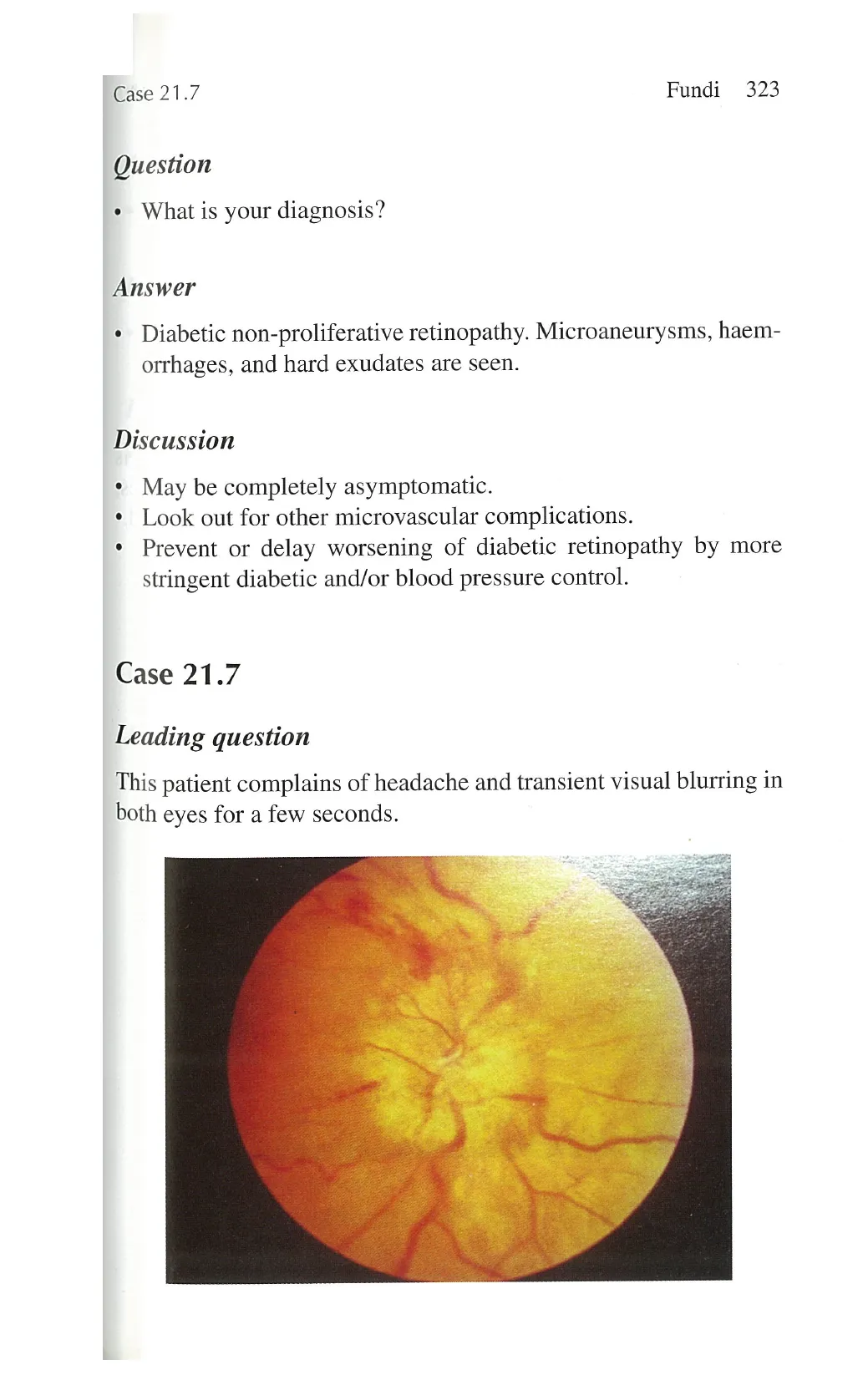
* Papilloedema.
Description
e Disc hyperemia, elevated with indistinct margins. There is
venous engorgement, peripapillary flame shaped haemorrhages,
cotton wool exudates. Visual acuity can be normal or reduced.
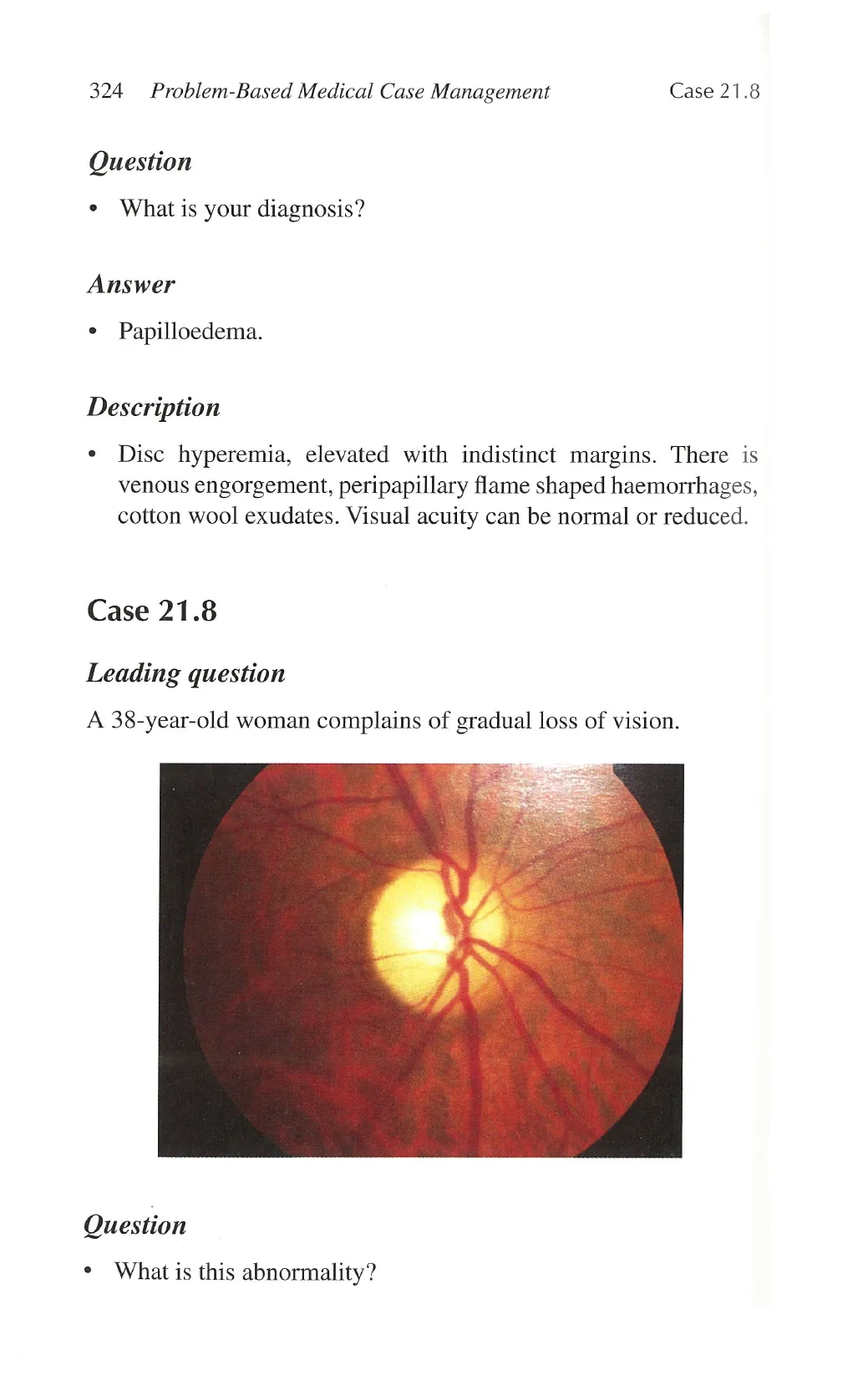
Case 21.8
Leading question
A 38-year-old woman complains of gradual loss of vision.
Question
* What is this abnormality?
View original page (figures / layout)
Page 9
Case 21.9 Fundi 325
Answer
e Primary optic atrophy.
Description
« White, flat disc with clear margins.
Discussion
¢ What are the common causes?
- Retrobulbar neuritis.
Compressive (tumours/aneurysms).
Hereditary optic neuropathies.
Toxic/nutritional optic neuropathies.
|
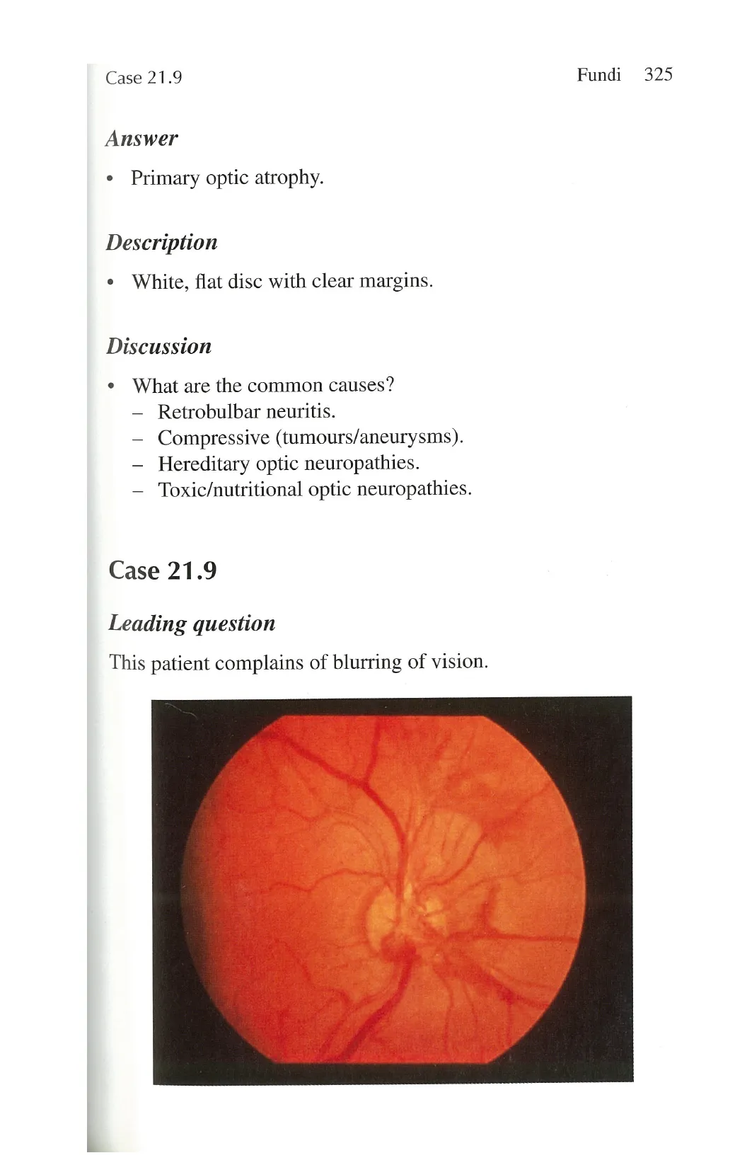
Case 21.9
Leading question
This patient complains of blurring of vision.
View original page (figures / layout)
Page 10
326 Problem-Based Medical Case Management Case 21.10
Question
What is your diagnosis?
Answer
¢ Proliferative diabetic retinopathy.
Description
e New blood vessel formation at 12-2 o’clock region, with blot
and dot haemorrhages at 4 o’clock.
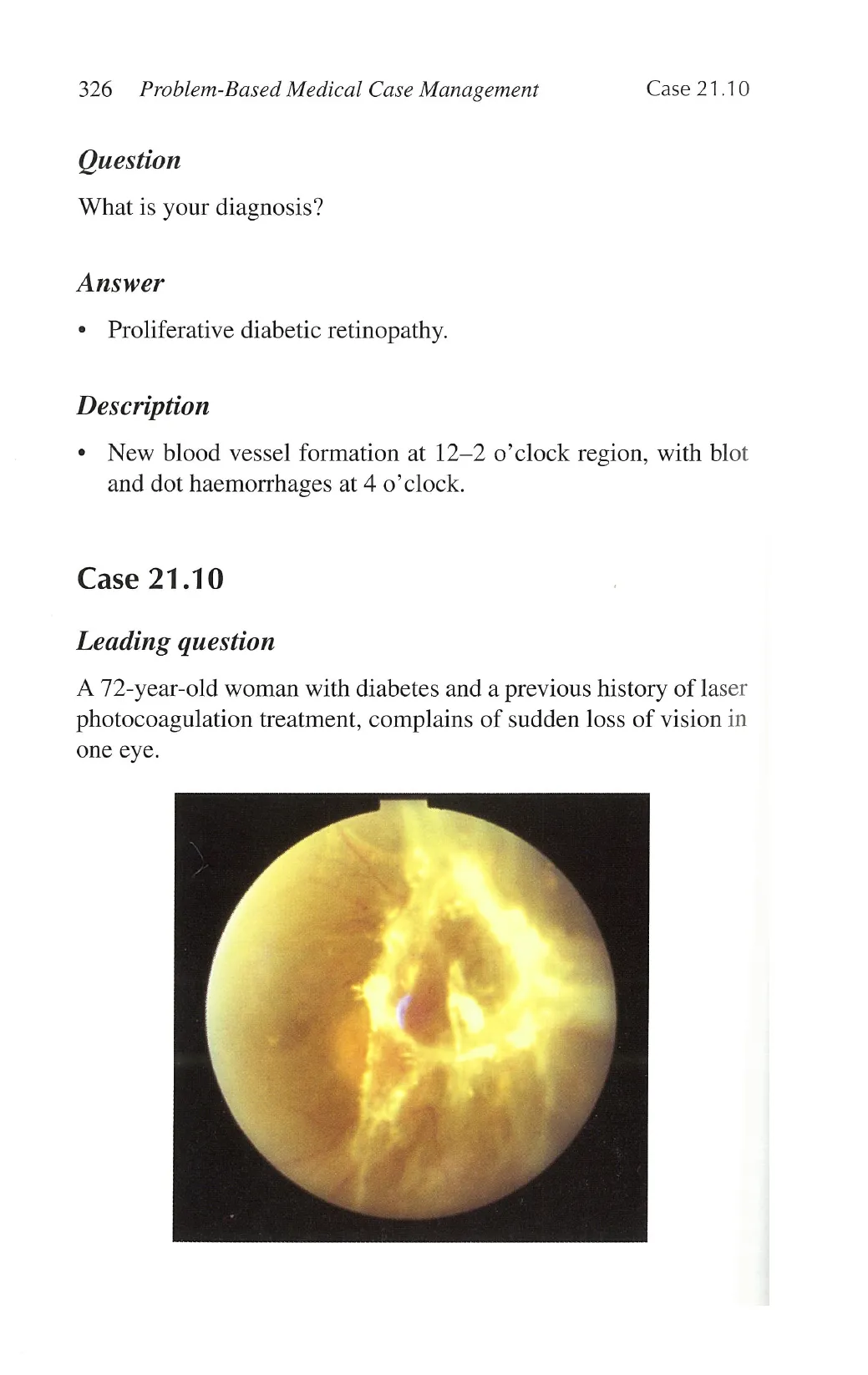
Case 21.10
Leading question
A 72-year-old woman with diabetes and a previous history of laser
photocoagulation treatment, complains of sudden loss of vision in
one eye.
View original page (figures / layout)
Page 11
Case 21.10 Fundi 327
Question
* What is the diagnosis?
Answer
+ Vitreous haemorrhage and tractional retinal detachment.
Discussion
* Healed vitreous haemorrhage predisposed to future risk of
retinal detachment.
View original page (figures / layout)