Page 1
20
Electrocardiogram
Hung-Fat Tse
Case 20.1
Leading question
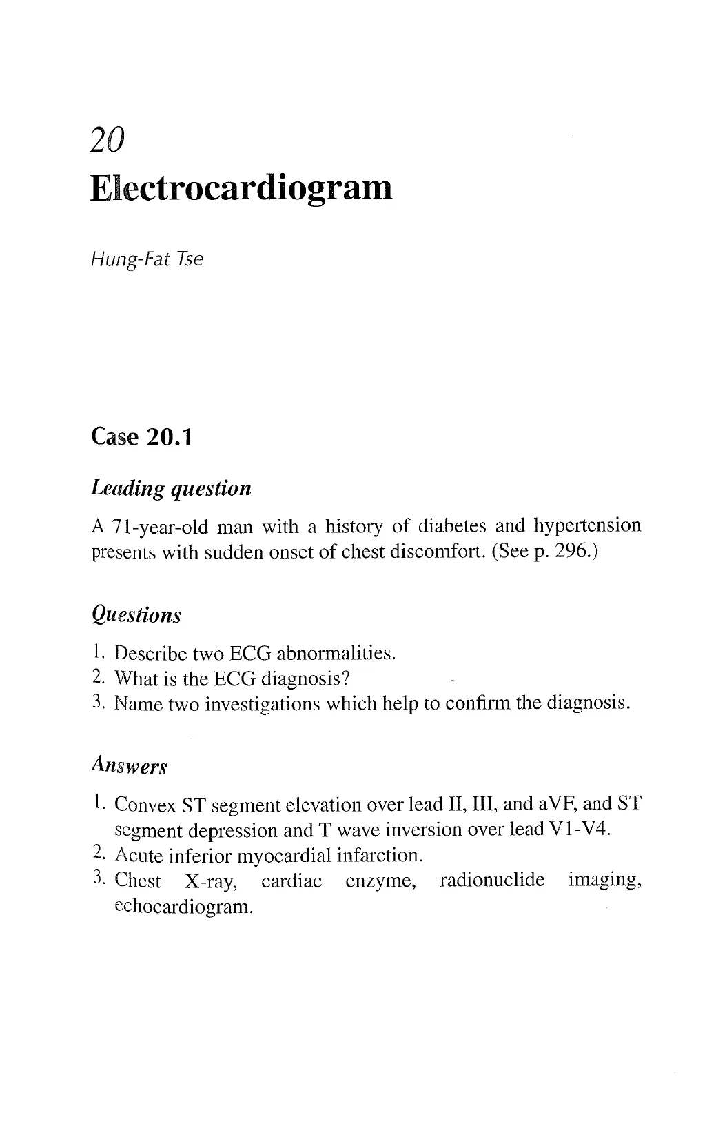
A 7l-year-old man with a history of diabetes and hypertension
presents with sudden onset of chest discomfort. (See p. 296.)
Questions
I. Describe two ECG abnormalities.
2. What is the ECG diagnosis?
3. Name two investigations which help to confirm the diagnosis.
Answers
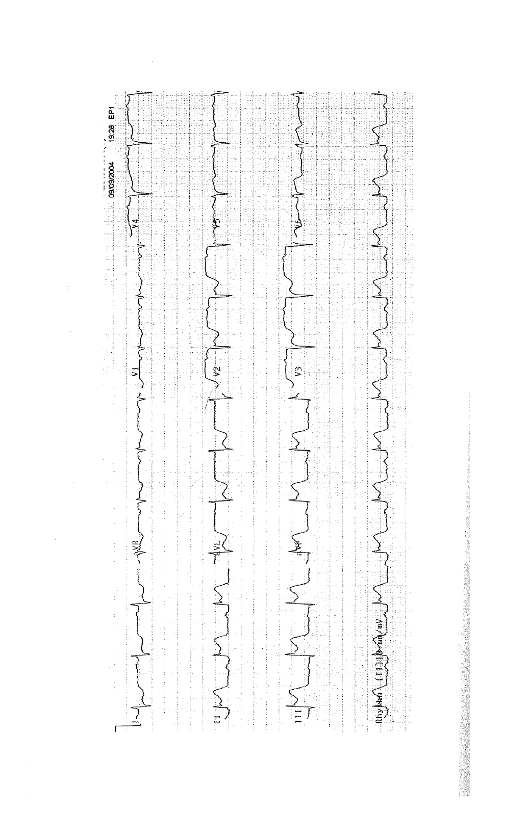
L. Convex ST segment elevation over lead IT, III, and aVF, and ST
segment depression and T wave inversion over lead V1-V4.
2. Acute inferior myocardial infarction.
3. Chest X-ray, cardiac enzyme, radionuclide imaging,
echocardiogram.
View original page (figures / layout)
Page 2
View original page (figures / layout)
Page 3
Case 20.1 Electrocardiogram 297
Description
+ The ECG abnormalities associated with acute myocardial
infarction typically evolve in a relatively predictable phase.
« The earliest ECG changes is marked peaking of the T wave—
“hyperacute T wave’—over the site of myocardial infarction.
If the myocardial ischemia persisted, the peaked T waves evolve
into ST segment elevation.
* Significant ST segment elevation is defined as = 1 mm
(0.10 mV) in 2 or more limb leads or precordial leads V4-V6
or = 2 mm (0.20 mV) in two or more precordial leads V1-V3.
The ST segment elevation of myocardial infarction is usually
upwardly convex.
* As the acute infarction continues to evolve, the ST segment
elevation decreases and the T wave begins to invert. As the ST
segment elevation subsides, the T wave usually becomes pro-
gressively deeper.
Discussion
* In contrast to convex ST segment elevation during acute myo-
cardial infarction, concave ST segment elevation is typically
seen in the setting of pericarditis or normal variant early repo-
larization abnormality.
* The potential clues to differentiate these conditions with acute
myocardial infarction includes:
~ The leads with ST segment elevation in acute infarction are
more localized than the diffuse changes in pericarditis (all
leads except aVR).
~ The presence of reciprocal ST depression over other leads
during acute myocardial infarction as shown in this case.
However, the ST segment changes over other leads can rep-
resent coexisting myocardial ischemia over other sites.
~ The presence of PR segment depression during acute peri-
carditis (rarely in acute myocardial infarction).
View original page (figures / layout)
Page 4
298 Problem-Based Medical Case Management Case 20.2
Case 20.2
Leading question
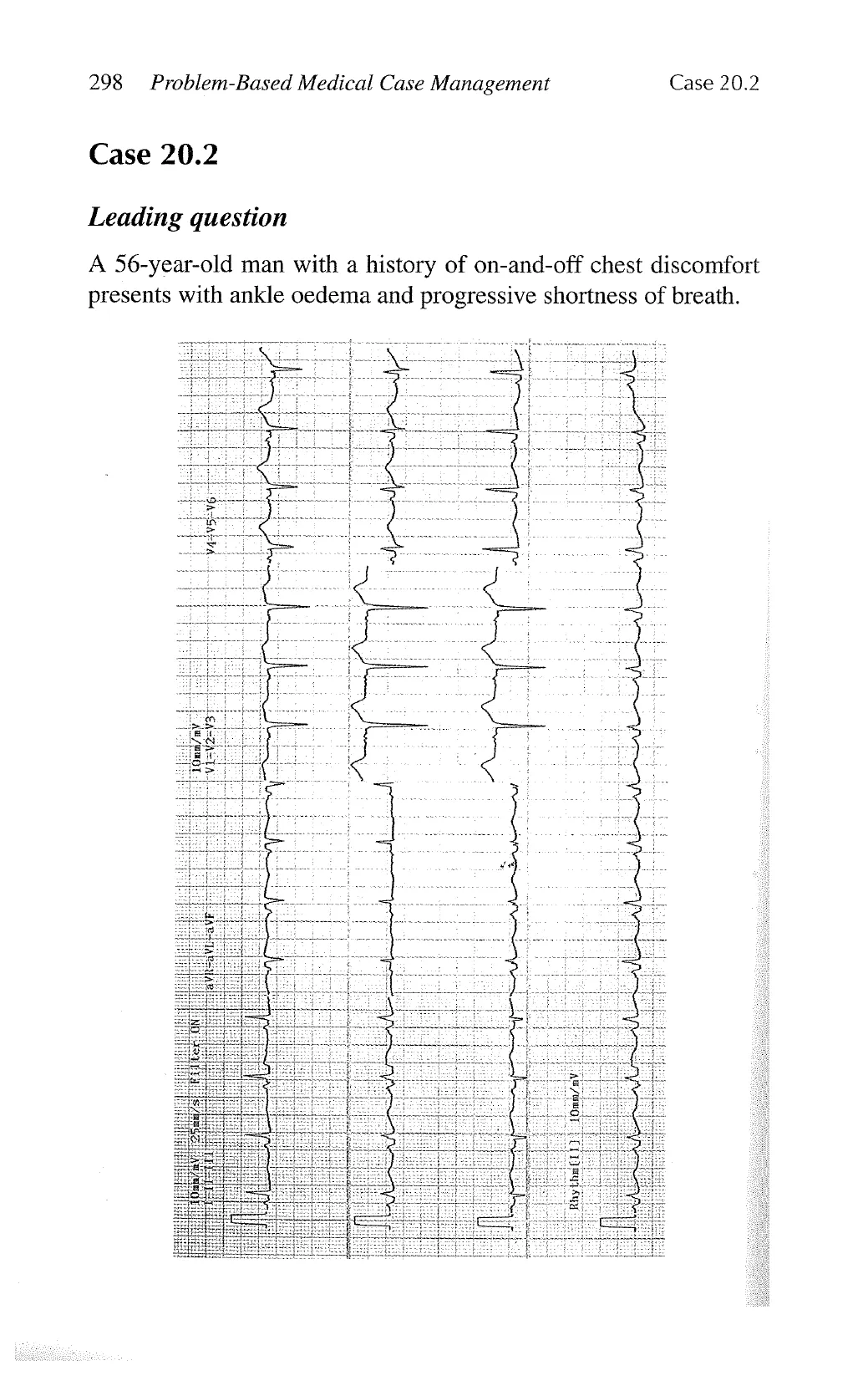
A 56-year-old man with a history of on-and-off chest discomfort
presents with ankle oedema and progressive shortness of breath.
View original page (figures / layout)
Page 5
Case 20.2 Electrocardiogram 299
Questions
1. Describe two ECG abnormalities.
2. What is the ECG diagnosis?
3. Name two investigations which help to confirm the diagnosis.
Answers
1. Pathological Q wave over V1-V3, and left atrial abnormality.
2. Old anteroseptal myocardial infarction.
3. Chest X-ray, cardiac enzyme, echocardiogram, radionuclide
imaging, cardiac catheterization, magnetic resonance imaging.
Description
+ Abnormal Q waves usually develop in the first several hours to
days after acute infarction following resolution of ST elevation.
In the majority of cases, Q waves persist indefinitely (~85%).
* Persistence of ST elevation with abnormal Q wave should raise
the suspicion of ventricular aneurysm.
* Q waves over lead III and aVR can be normal.
* The presence of isoelectric ST segments and upright T waves
suggest that the infarction is not acute (months to years).
Discussion
* Other than myocardial infarction, abnormal Q wave can be
observed in the following conditions:
~ Wolff-Parkinson-White syndrome: negative delta-waves
over inferior lead can mimic inferior myocardial infarction.
~ Left bundle branch block: QS pattern over V1-V4 together
with 1-2 mm ST segment elevation mimic anteroseptal
myocardial infarction.
— Chronic obstructive airway obstruction: Q wave over infe-
rior lead with poor R wave progression, P pulmonale, and
low voltage QRS.
View original page (figures / layout)
Page 6
300 Problem-Based Medical Case Management Case 20.3
— Hypertrophic cardiomyopathy: septal hypertrophy with Q
wave over I, aVL, V4-V6.
— Pulmonary embolism: Q wave over lead III and aVF associ-
ated with ST and T wave changes can mimic inferior myo-
cardial infarction, but Q wave over lead II is rare.
Case 20.3
Leading question
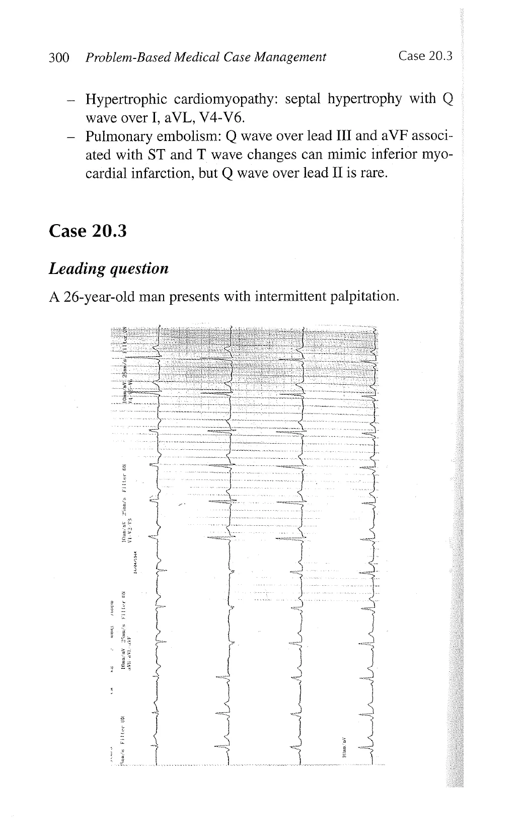
A 26-year-old man presents with intermittent palpitation.
3
i
J
: ¥ ! Y
i J
L (SRS —
AJ
[
L
}
View original page (figures / layout)
Page 7
Case 20.3 Electrocardiogram 301
Questions
1. Describe two ECG abnormalities.
2. What is the ECG diagnosis?
3. Name two abnormal cardiac arrhythmias that can be associated
with this condition.
Answers
1. Short PR interval, and presence of delta wave.
2. Wolff-Parkinson-White syndrome.
3. Atrial fibrillation, ventricular fibrillation, paroxysmal supraven-
tricular tachycardia.
Description
* The presence of a short PR interval (< 0.12 s), wide QRS
complex (> 0.1 s), delta wave (prolonged upstroke of QRS
complex) with or without secondary ST and T wave changes
indicative of Wolff-Parkinson-White syndrome.
Discussion
* Other causes of an RSR’ complex in V1 include:
~ Right ventricular hypertrophy: associated with right axis
deviation, right atrial enlargement, R wave > 10 mm and ST
depression and T wave inversion over V1-V3.
— Posterior myocardial infarction: associated with inferior
myocardial infarction.
~ Right bundle branch block: QRS duration > 120 ms, T wave
inversion over V1-V2 and wide S wave in V5-V6.
View original page (figures / layout)
Page 8
302 Problem-Based Medical Case Management Case 20.4
Case 20.4
Leading question
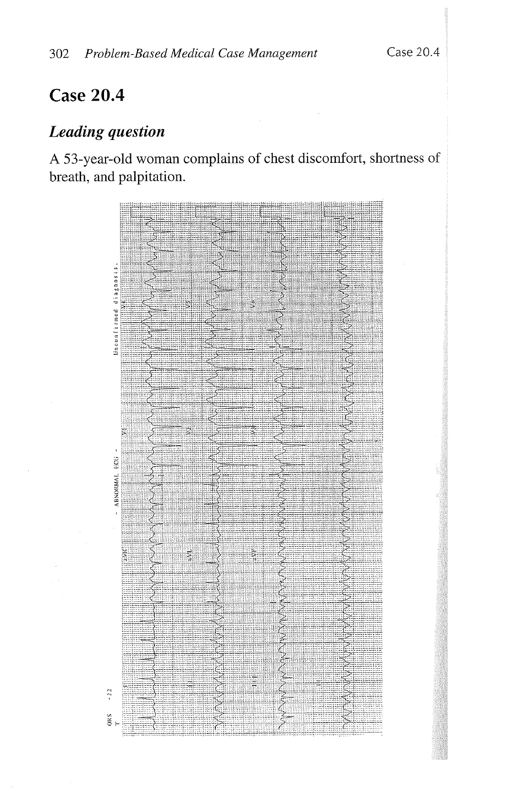
A 53-year-old woman complains of chest discomfort, shortness of
breath, and palpitation.
View original page (figures / layout)
Page 9
Case 20.4 Electrocardiogram 303
Questions
1. Describe two ECG abnormalities.
2. What is the ECG diagnosis?
3. Name two appropriate investigations.
4, Name two drugs that can control the heart rate and relieve her
symptoms.
Answers
1. Narrow complex tachycardia (rate 150 bpm) and flutter wave.
2. Atrial flutter with 2:1 conduction.
3. Chest X-ray, electrolytes, cardiac enzymes, thyroid function
test, echocardiogram.
4. Digoxin, diltiazem, verapamail, beta-blockers, and amiodarone.
Description
* Atrial flutter shows rapid regular atrial undulation (flutter
waves) at a rate of 240-340 per minute.
* Atrioventricular conduction ratio is usually fixed (2:1, 3:1 or >
4:1), but may be variable and mimic atrial fibrillation.
Discussion
* The response to carotid sinus massage or drugs (e.g., adeno-
sine or ATP) which decrease atrioventricular conduction can
unmark the atrial activity during atrial flutter. In contrast, these
manoeuvers terminate other regular supraventricular tachycar-
dias (atrioventricular nodal re-entry or atrioventricular re-entry
tachycardias).
View original page (figures / layout)
Page 10
304 Problem-Based Medical Case Management Case 20.5
Case 20.5
Leading question
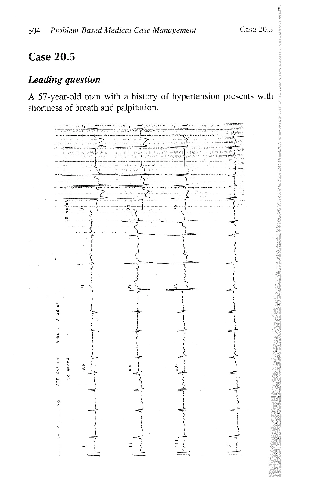
A 57-year-old man with a history of hypertension presents with
shortness of breath and palpitation.
Sokol. 3.32 au
18 mn/mV
aur
Coeoem /oo ks 0TC 433 as
1
X
I
View original page (figures / layout)
Page 11
Case 20.5 Electrocardiogram 305
Questions
1. Name two ECG diagnoses.
2. Suggest two appropriate investigations.
3. Name two drugs that can control the heart rate and relieve his
symptoms.
Answers
1. Atrial fibrillation and left ventricular hypertrophy.
2. Chest X-ray, electrolytes, cardiac enzymes, thyroid function
test, echocardiogram.
3. Digoxin, diltiazem, verapamail, beta-blockers, and amiodarone.
Description
+ Discrete P waves are absent during atrial fibrillation and atrial
activity is totally irregular and manifest as fibrillatory rate wave
with varying amplitudes, duration, and morphology. During
atrial fibrillation, ventricular rhythm is irregularly irregular
(unless the presence of complete heart block).
* ECG diagnosis of left ventricular hypertrophy is based on the
presence of left amplitude QRS complexes (S in V1 + Rin V5
or V6 > 35 mm or any R + S >45 mm).
* Down-sloping ST depression and asymmetrical T wave inver-
sion: “strain pattern” are present.
Discussion
* Atrial fibrillation may be mimicked by:
- Mu;El/i,focal atrial tachycardia-atrial rate > 100 bpm with 3 or
more different P wave morphologies, and PR, RR, and RP
intervals vary.
— Atrial flutter or atrial tachycardia with variable block—rapid
and regular P wave or flutter wave is observed.
* Digoxin toxicity may result in regularization of the RR inter-
val due to complete heart block with junctional tachycardia.
View original page (figures / layout)
Page 12
306 Problem-Based Medical Case Management Case 20.6
It can be exacerbated by hypokalaemia, hypomagnesaemia, and
hypercalcaemia.
* ST segment depression and T wave inversion can also be
observed with digoxin therapy and myocardial ischemia.
Case 20.6
Leading question
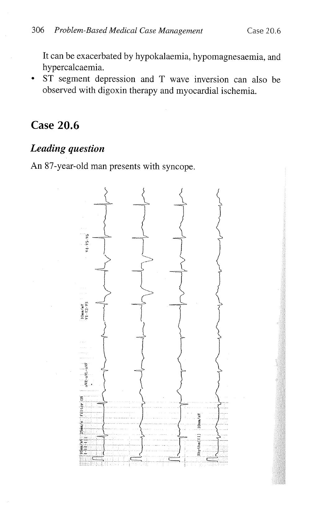
An 87-year-old man presents with syncope.
10nn/aV.
VI-v2-V3
)
Rbythalil) | 10mn/ay
View original page (figures / layout)
Page 13
Case 20.6 Electrocardiogram 307
Questions
1. Name three ECG abnormalities.
2. What is the ECG diagnosis?
3. Suggest two appropriate investigations.
4. What is the immediate treatment for this patient?
Answers
1. Right bundle branch block, left axis deviation, bradycardia,
atrioventricular dissociation.
. Complete heart block.
3. Chest X-ray, electrolytes, cardiac enzymes, thyroid function
test.
4. Temporary external or transvenous cardiac pacing.
)
Description
* During complete heart block, the atrial and ventricular activities
are independent of each other, unually described as atrioven-
tricular dissociation. As a result, the PP and RR intervals are
constant, but the PR interval varies.
* Furthermore, the atrial rate is faster than the ventricular rate
during complete heart block.
Discussion
* In the majority of cases, complete heart block is caused by
degeneration of conduction system, and rarely due to congeni-
tal heart block.
* Potential reversible causes of complete heart block include
hyperkalaemia, endocarditis, acute myocardial infarction,
digoxin toxicity, and Lyme diseases.
View original page (figures / layout)
Page 14
308 Problem-Based Medical Case Management Case 20.7
Case 20.7
Leading question
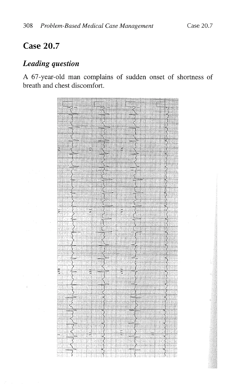
A 67-year-old man complains of sudden onset of shortness of
breath and chest discomfort.
View original page (figures / layout)
Page 15
Case 20.7 Electrocardiogram 309
Questions
. Name two ECG abnormalities.
. What is the diagnosis?
. Suggest two appropriate investigations to confirm the diagnosis.
W oo —
Answers
1. Sinus tachycardia and S1Q3T3.
2. Pulmonary embolism.
3. Chest X-ray, CT scan of thorax, ventilation-perfusion scan, and
pulmonary angiogram.
Description
* During acute pulmonary embolism with acute cor pulmonale,
changes appear in the frontal plane leads (I, IIT, aVF, V2-V3)
that mimic acute inferior myocardial infarction.
* Lead IIT is mainly involved with an increase in the size of Q
wave, slight ST segment elevation, and shallow inversion of the
T wave (Q3T3).
* Other changes include increase size of S wave in lead I (S1),
right axis deviation, right bundle branch block, and sinus
tachycardia.
Discussion
* The presence of sinus tachycardia and findings consistent with
right ventricular pressure overload as described above are sug-
gestive of acute pulmonary embolism.
*+ ECG abnormalities of pulmonary embolism are often transient.
* Other causes of right ventricular overload include right ven-
tricular hypertrophy with pulmonary hypertension and chronic
obstructive airway diseases.
View original page (figures / layout)
Page 16
310 Problem-Based Medical Case Management Case 20.8
Case 20.8
Leading question
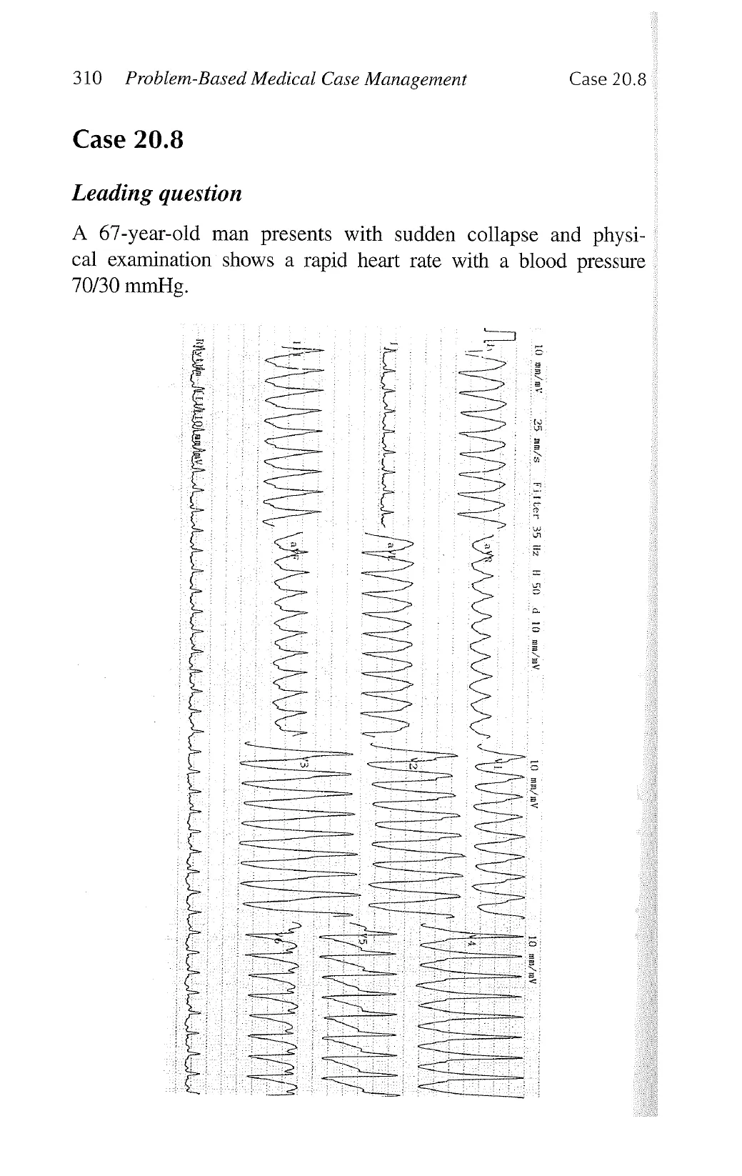
A 67-year-old man presents with sudden collapse and physi-
cal examination shows a rapid heart rate with a blood pressure
70/30 mmHg.
1
A gy
{
s/ 7
05 07 SE i
Awun gy p
Auju o1
Al g1
View original page (figures / layout)
Page 17
Case 20.8 Electrocardiogram 311
Questions
|. What is the ECG diagnosis?
. List three possible causes for this ECG abnormality.
. Suggest two appropriate investigations.
. What is the immediate treatment for this patient?
FNIES)
Answers
1. Wide complex tachycardia.
2. Ventricular tachycardia, supraventricular tachycardia with pre-
existing right bundle branch block, supraventricular tachycardia
with aberrant conduction and antidromic tachycardia through
accessory pathway.
. Chest X-ray, electrolytes, cardiac enzymes, echocardiogram.
4. External DC cardioversion.
>
Description
* QRS morphology during wide complex tachycardia that favours
ventricular tachycardia include atrioventricular dissociation,
capture beats, fusion beats, > 140 ms for right bundle branch
block pattern or > 160 ms for left bundle branch block pattern,
when QRS deflections in the precordial leads are all positive or
negative (concordance) and when the R wave is taller than the
R’ wave in V1.
Discussion
* In contrast, ECG features suggestive of supraventricular tachy-
cardia with aberrancy include QRS morphology similar to sinus
rthythm or aberrantly conducted atrial premature beats, QRS
deflection discordance in precordial lead.
* All haemodynamic unstable wide complex tachycardia should
be treated as ventricular tachycardia with DC cardioversion.
View original page (figures / layout)
Page 18
312 Problem-Based Medical Case Management Case 20.9
Case 20.9
Leading question
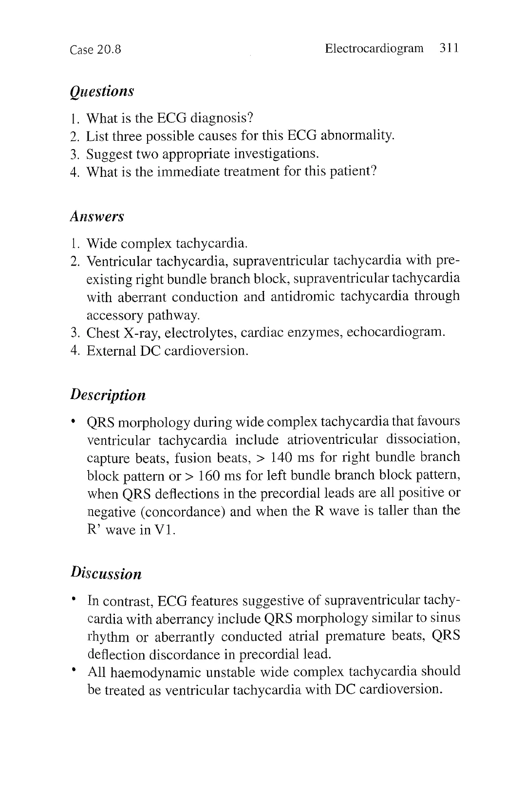
A 52-year-old man presents with sudden onset of palpitation.
1
Ty
|
i
0000-00000 507
sasfus s2ipasds
Aufwu gytisany Awfws 01qui
View original page (figures / layout)
Page 19
Case 20.9 Electrocardiogram 313
Questions
I
2
3
4
5
. Name three ECG abnormalities.
. What is the ECG diagnosis?
. What are the possible mechanisms for this ECG diagnosis?
. Suggest two appropriate investigations.
. What is the immediate treatment for this patient?
Answers
1.
w o
Narrow complex tachycardia, retrograde atrial activation, and
ST and T wave abnormalities.
. Supraventricular tachycardia.
. Atrioventricular nodal re-entry tachycardia, atrioventricular
re-entry tachycardia, and atrial tachycardia.
. Chest X-ray, electrolytes, cardiac enzymes, thyroid function
test.
. Vagal manoeuvers, fast-acting atrioventricular nodal blocking
agents, such as adenosine and verapamail.
Description
During supraventricular tachycardia, the heart rate is > 100 bpm,
the QRS complex is regular and narrow, and the P wave is not
easily identified.
Retrograde atrial activation lead to inverted P waves in leads II,
III, and aVE.
ST and T wave abnormalities suggestive of myocardial ischemia
are common during rapid tachycardia.
Discussion
Although the number and location of P wave may provide a cue
to the diagnosis of the mechanisms of supraventricular tachy-
cardia, it is difficult to distinguish them based solely on ECG:
~ Atrioventricular nodal re-entry tachycardia: P wave fused to
or immediately after QRS complex.
View original page (figures / layout)
Page 20
314 ' Problem-Based Medical Case Management Case 20.10
— Atrioventricular re-entry tachycardia: P wave in the ST
segment.
— Atrial tachycardia: P wave > QRS complex.
Case 20.10
Leading question
A 67-year-old woman presents with syncope after taking medica-
tions for “flu” symptoms.
NN
NN
N
o~ dd~
L~
i
I
I
|
s
!
\J‘
e
!
SN
[N
Tty
o8
AT PA G I
|
%
e
)
-
View original page (figures / layout)
Page 21
Case 20.10 Electrocardiogram 315
Questions
1. Name three ECG abnormalities.
2. List three possible causes for this ECG abnormality.
3. Suggest two appropriate investigations.
Answers
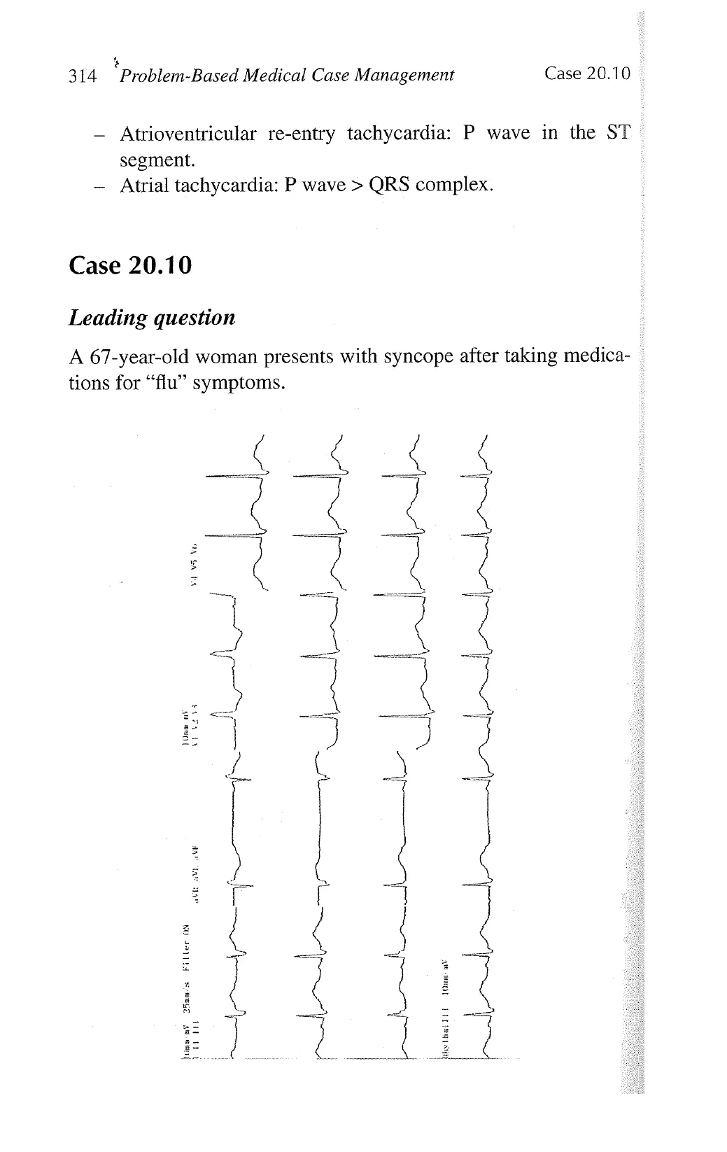
1. Sinus bradycardia, right bundle branch block, sinus arrhythmia,
and prolonged QT interval.
2. The possible causes are:
- Drugs: anti-arrhythmic agents, antibiotic (erythromycin,
clarithromycin), antihistamine (terfenedine, diphenhy-
dramine), and anti-pyschotic agents.
- Electrolytes imbalance: hypokalaemia, hypocalcaemia, and
hypomagnesaemia.
- Congenital long QT syndrome.
— Metabolic disorder: hypothyroidism and diabetes.
— Neurologic disorder: ~stroke, trauma, subarachnoid
haemorrhage.
— TIschemic heart disease and bradycardia.
3. Electrolytes, cardiac enzymes, thyroid function test.
Description
* Normal QT interval varies inversely with heart rate. Therefore,
measurement of QT interval needs to be corrected with heart
rate: QTc = QT/YRR interval (in second).
* During right bundle branch block, the QRS duration > 120 ms,
RSR’ pattern with R’ taller than R wave, wide slurred S wave in
lead V5-V6, and ST segment depression and T wave inversion
over V1-V2.
Discussion
* Prolonged QT interval is associated with polymorphic ventricu-
lar tachycardia—torsades de pointes.
View original page (figures / layout)
Page 22
316 Problem-Based Medical Case Management Case 20.10
* In contrast to left bundle branch block, right bundle branch
block can occur in normal subjects, and does not interfere with
ECG diagnosis of left ventricular hypertrophy or ST elevation
myocardial infarction.
View original page (figures / layout)