Page 1
19
Respiratory Diagnostics
| David Chi-Leung Lam, Christopher Kim-Ming Hui,
| Jamie Chung-Mei Lam, Julie Kwan-Ling Wang,
| and Macy Mei-Sze Lui
19.1 CXRs and CT thorax images
Case 1
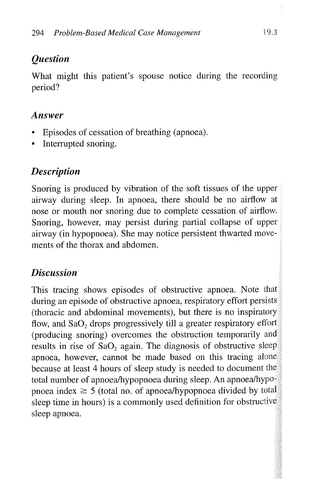
A 56-year-old man presented with fever and cough with the fol-
lowing chest X-ray.
What is the radiological sign indicated by the arrows?
Answer: Air bronchogram.
View original page (figures / layout)
Page 2
270 Problem-Based Medical Case Management 19.1
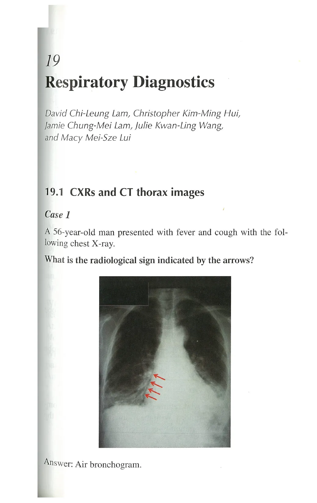
Below is the CT thorax of the same patient
5. 0y
What is the name of this CT window?
Answer: Lung window.
Which lobes are most severely affected?
Answer: The right and the left lower lobes, with the left lower lobe
more extensively affected than the right lower lobe.
What is the clinical diagnosis?
Answer; Community-acquired pneumonia (bilateral lower lobe
pneumonia). .
Case 2
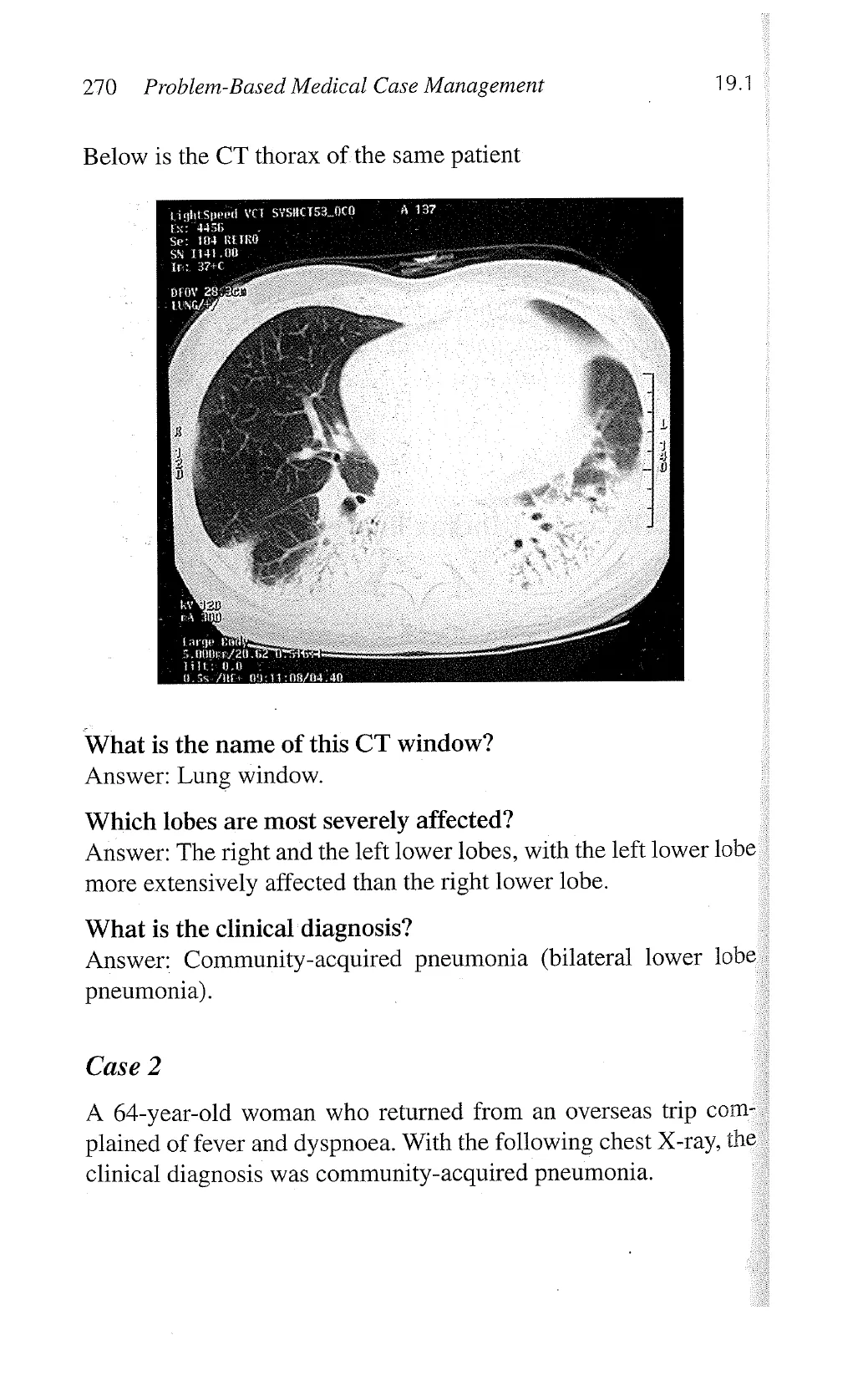
A 64-year-old woman who returned from an overseas trip com- .
plained of fever and dyspnoea. With the following chest X-ray, the
clinical diagnosis was community-acquired pneumonia.
View original page (figures / layout)
Page 3
19.1 Respiratory Diagnostics 271
Which lobe(s) of the lungs was/were involved?
Answer: The right middle lobe and right lower lobe.
The CXR shows right lower lobe consolidation and right middle
lobe consolidation-collapse.
The patient was suspected to have pneumonia. Suggest which
anatomical description best fits this type of pneumonia.
Answer: Multilobar pneumonia.
Which are the common pathogens isolated in community-
acquired pneumonia?
Answer: Streptococcus pneumoniae, Hemophilus influenza,
Moraxella catarrhalis, and Staphylococcus aureus.
What are the tests for identification of these microbiologic
pathogens?
Answer:
* Bacterial smear and culture of airway specimens (sputum or
bronchial lavage).
Blood culture for bacteria.
“Atypical pneumonia” caused by Legionella pneumophila,
Mycoplasma pneumoniae or Chlamydia psittacci are not
View original page (figures / layout)
Page 4
272 Problem-Based Medical Case Management 19.1
considered “isolates” as these microorganisms are difficult
to be cultured and the diagnosis rely on clinical presentation,
acute, and convalescent antibody titres, or urine for legionella
antigen. In some laboratories, PCR tests for the microbes are
available.
Case 3
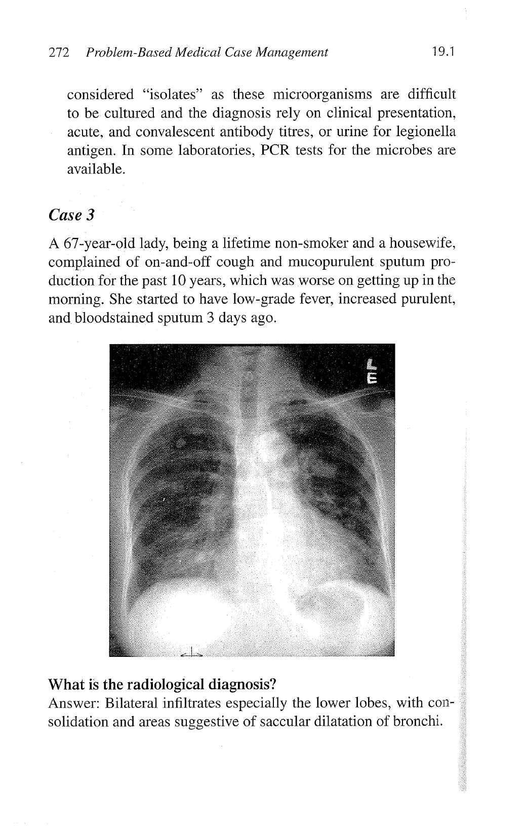
A 67-year-old lady, being a lifetime non-smoker and a housewife,
complained of on-and-off cough and mucopurulent sputum pro-
duction for the past 10 years, which was worse on getting up in the
morning. She started to have low-grade fever, increased purulent,
and bloodstained sputum 3 days ago.
What is the radiological diagnosis?
Answer: Bilateral infiltrates especially the lower lobes, with con- -
solidation and areas suggestive of saccular dilatation of bronchi.
View original page (figures / layout)
Page 5
19.1 Respiratory Diagnostics 273
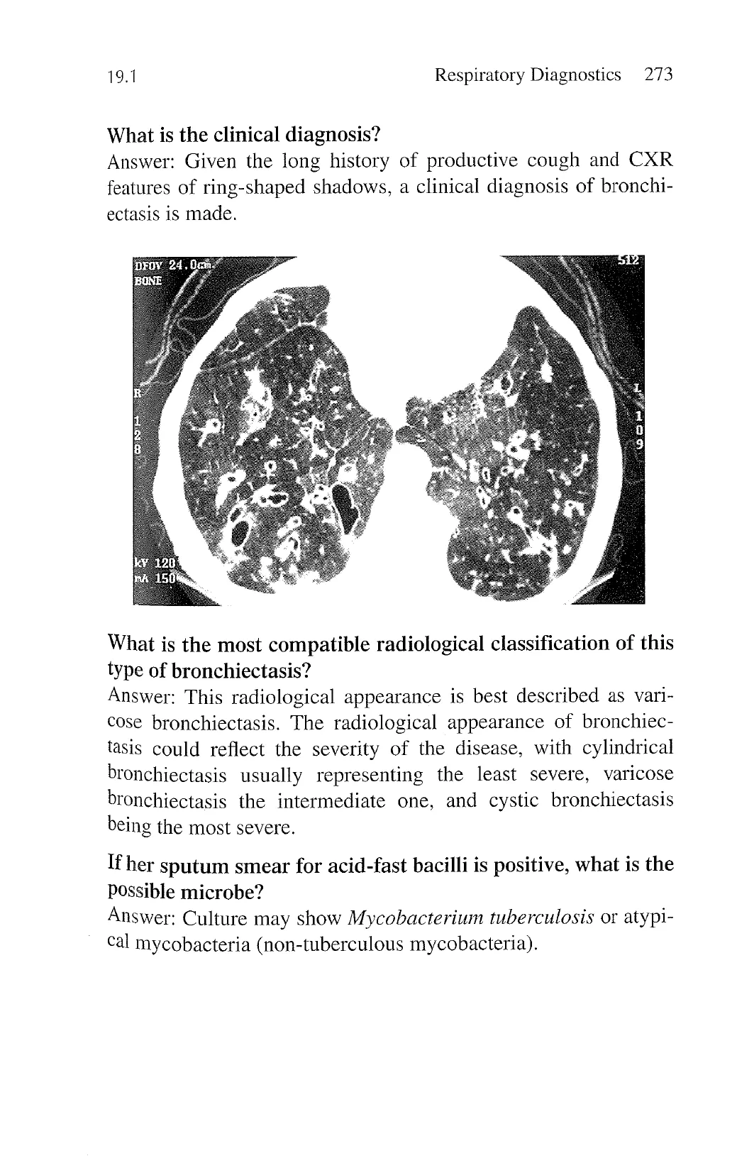
What is the clinical diagnosis?
Answer: Given the long history of productive cough and CXR
features of ring-shaped shadows, a clinical diagnosis of bronchi-
ectasis is made.
What is the most compatible radiological classification of this
type of bronchiectasis?
Answer: This radiological appearance is best described as vari-
cose bronchiectasis. The radiological appearance of bronchiec-
tasis could reflect the severity of the disease, with cylindrical
bronchiectasis usually representing the least severe, varicose
bronchiectasis the intermediate one, and cystic bronchiectasis
being the most severe.
If her sputum smear for acid-fast bacilli is positive, what is the
Ppossible microbe?
Answer: Culture may show Mycobacterium tuberculosis or atypi-
cal mycobacteria (non-tuberculous mycobacteria).
View original page (figures / layout)
Page 6
274 Problem-Based Medical Case Management 19.1
Case 4
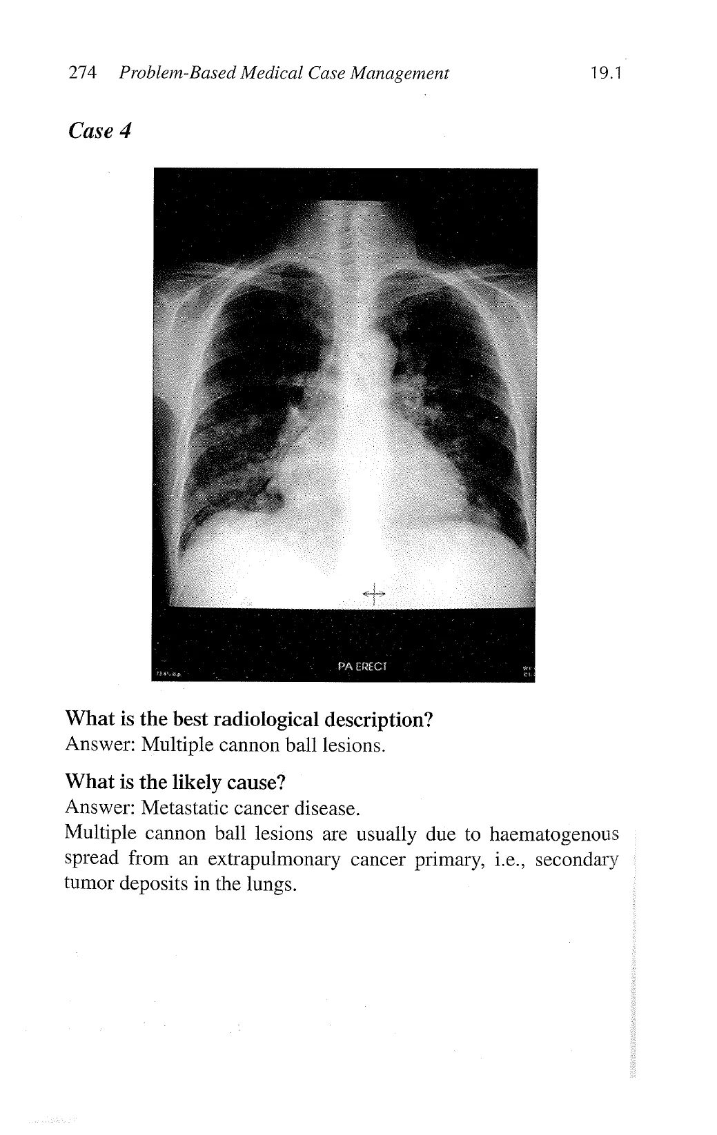
What is the best radiological description?
Answer: Multiple cannon ball lesions.
What is the likely cause?
Answer: Metastatic cancer disease.
Multiple cannon ball lesions are usually due to haematogenous
spread from an extrapulmonary cancer primary, i.e., secondary
tumor deposits in the lungs.
View original page (figures / layout)
Page 7
19.1 Respiratory Diagnostics 275
Case 5
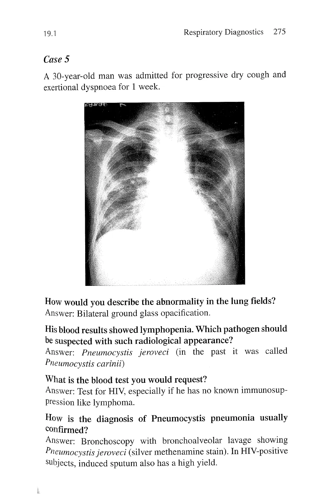
A 30-year-old man was admitted for progressive dry cough and
exertional dyspnoea for 1 week.
How would you describe the abnormality in the lung fields?
Answer: Bilateral ground glass opacification.
His blood results showed lymphopenia. Which pathogen should
be suspected with such radiological appearance?
Answer: Pneumocystis jeroveci (in the past it was called
Pneumocystis carinii)
What is the blood test you would request?
Answer: Test for HIV, especially if he has no known immunosup-
Pression like lymphoma.
How is the diagnosis of Pneumocystis pneumonia usually
confirmed?
Answer: Bronchoscopy with bronchoalveolar lavage showing
Pneumocystis jeroveci (silver methenamine stain). In HIV-positive
Subjects, induced sputum also has a high yield.
View original page (figures / layout)
Page 8
276 Problem-Based Medical Case Management 19.1
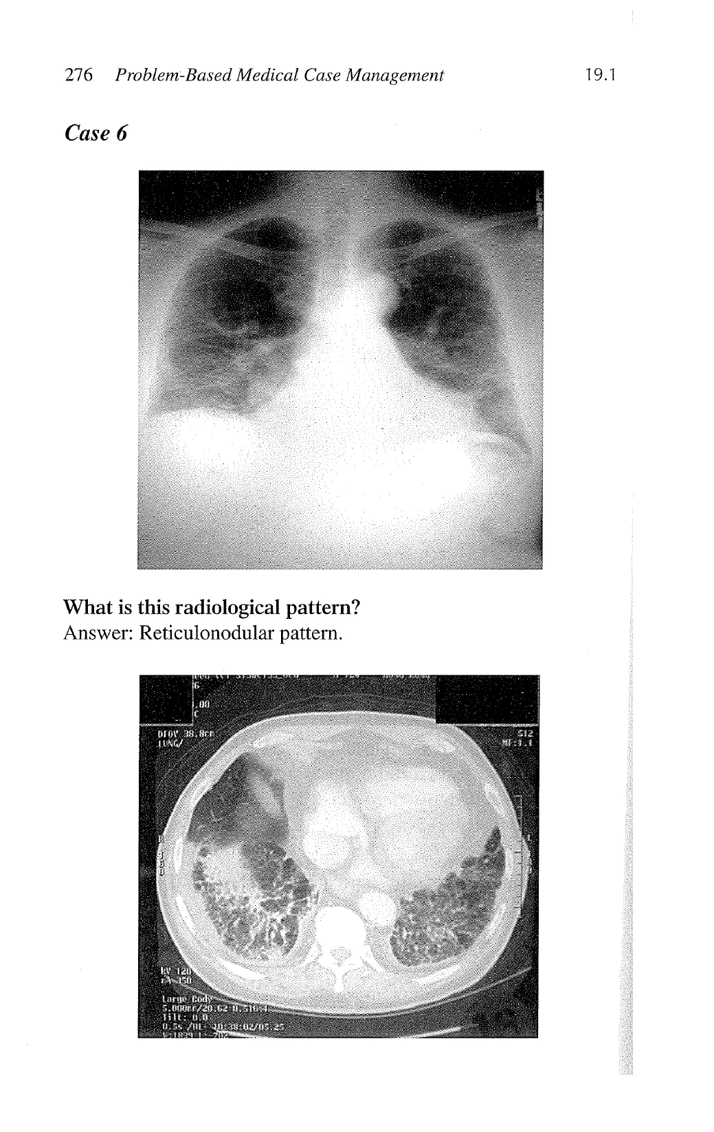
Case 6
What is this radiological pattern?
Answer: Reticulonodular pattern.
.00
‘,
8 er
View original page (figures / layout)
Page 9
19.1 Respiratory Diagnostics 277
What is the best radiological description?
Answer: Honeycombing, characteristic of interstitial pulmonary
fibrosis.
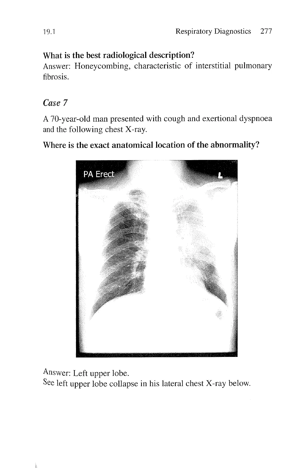
Case 7
A 70-year-old man presented with cough and exertional dyspnoea
and the following chest X-ray.
Where is the exact anatomical location of the abnormality?
PA Erect
Answer: Left upper lobe.
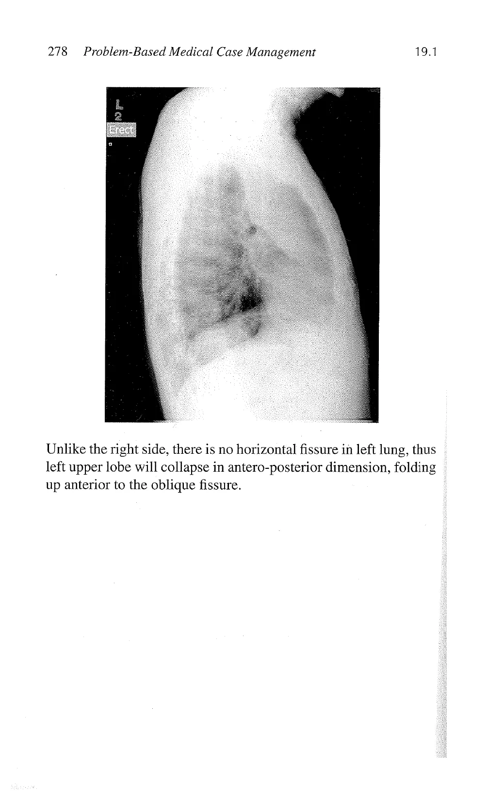
See left upper lobe collapse in his lateral chest X-ray below.
View original page (figures / layout)
Page 10
278 Problem-Based Medical Case Management 19.1
Unlike the right side, there is no horizontal fissure in left lung, thus
left upper lobe will collapse in antero-posterior dimension, folding
up anterior to the oblique fissure.
View original page (figures / layout)
Page 11
19.1 Respiratory Diagnostics 279
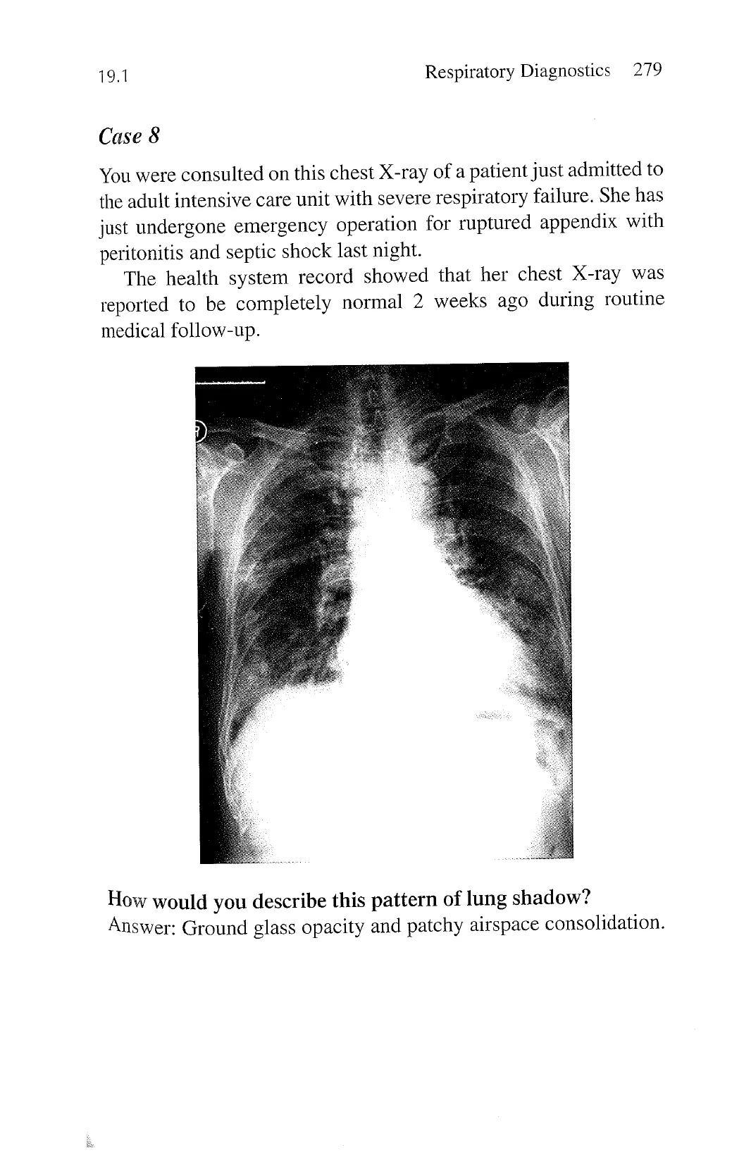
Case 8
You were consulted on this chest X-ray of a patient just admitted to
the adult intensive care unit with severe respiratory failure. She has
just undergone emergency operation for ruptured appendix with
peritonitis and septic shock last night.
The health system record showed that her chest X-ray was
reported to be completely normal 2 weeks ago during routine
medical follow-up.
How would you describe this pattern of lung shadow?
Answer: Ground glass opacity and patchy airspace consolidation.
View original page (figures / layout)
Page 12
280 Problem-Based Medical Case Management 19.1
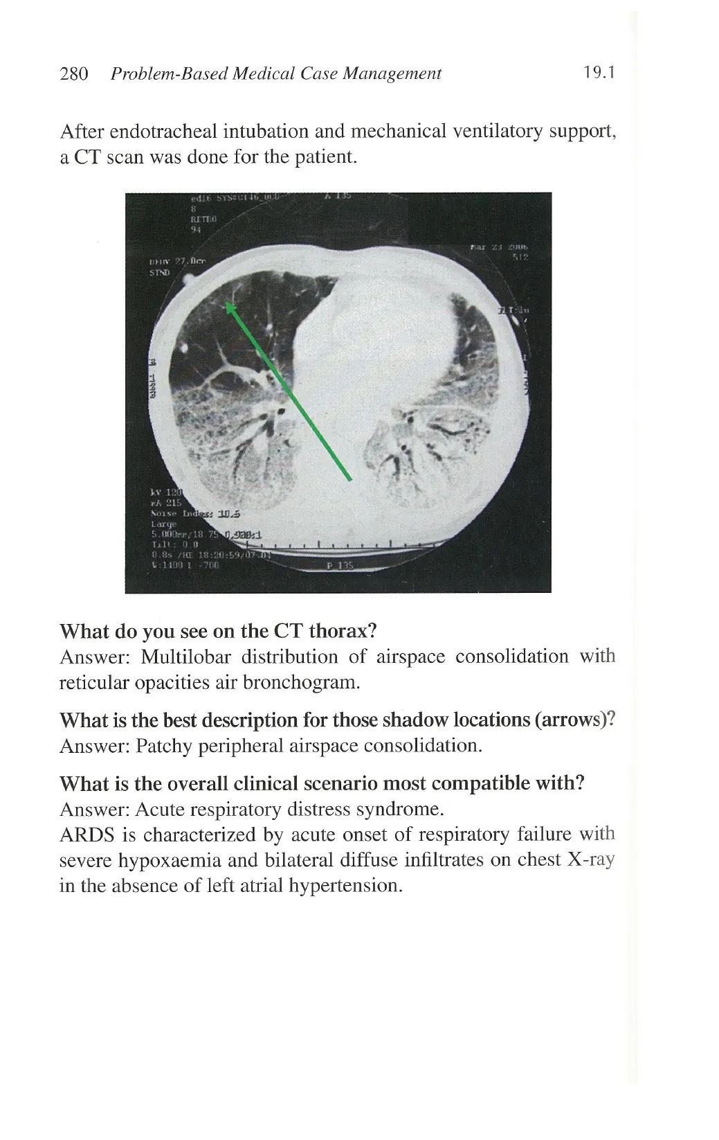
After endotracheal intubation and mechanical ventilatory support,
a CT scan was done for the patient.
What do you see on the CT thorax?
Answer: Multilobar distribution of airspace consolidation with
reticular opacities air bronchogram.
What is the best description for those shadow locations (arrows)?
Answer: Patchy peripheral airspace consolidation.
What is the overall clinical scenario most compatible with?
Answer: Acute respiratory distress syndrome.
ARDS is characterized by acute onset of respiratory failure with
severe hypoxaemia and bilateral diffuse infiltrates on chest X-ray
in the absence of left atrial hypertension.
View original page (figures / layout)
Page 13
19.1 Respiratory Diagnostics 281
Case 9
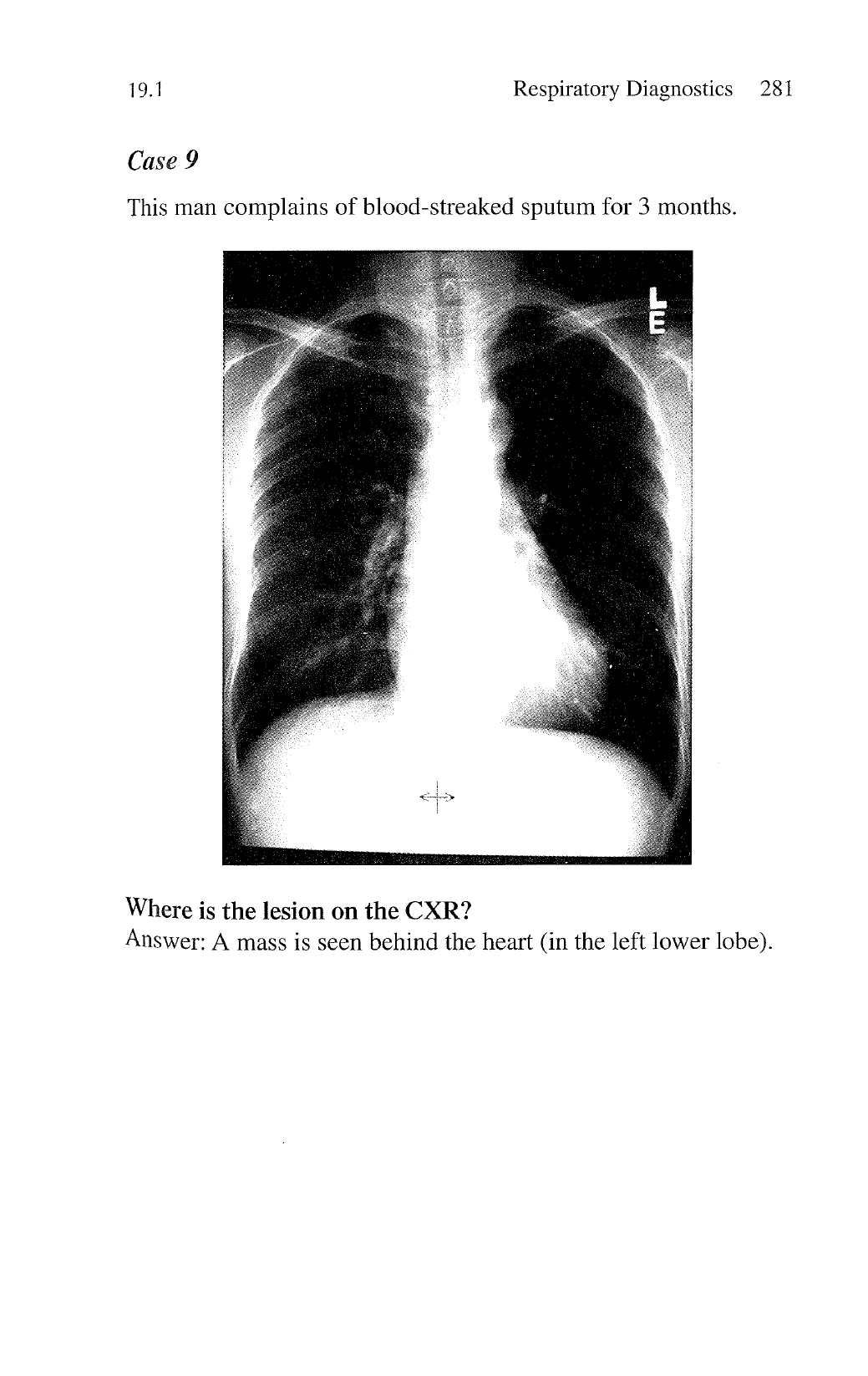
This man complains of blood-streaked sputum for 3 months.
Where is the lesion on the CXR?
Answer: A mass is seen behind the heart (in the left lower lobe).
View original page (figures / layout)
Page 14
282 Problem-Based Medical Case Management 19.1
Case 10
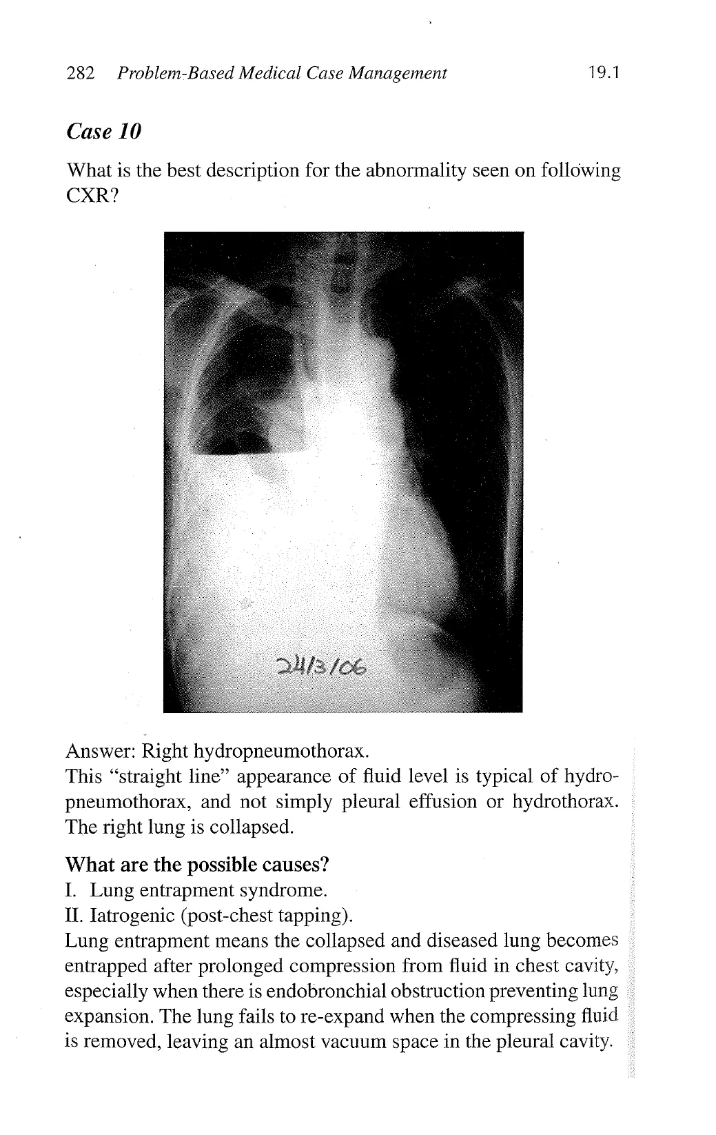
What is the best description for the abnormality seen on following
CXR?
Answer: Right hydropneumothorax.
This “straight line” appearance of fluid level is typical of hydro-
pneumothorax, and not simply pleural effusion or hydrothorax.
The right lung is collapsed.
What are the possible causes?
I. Lung entrapment syndrome.
1L Iatrogenic (post-chest tapping).
Lung entrapment means the collapsed and diseased lung becomes
entrapped after prolonged compression from fluid in chest cavity,
especially when there is endobronchial obstruction preventing lung
expansion. The lung fails to re-expand when the compressing fluid
is removed, leaving an almost vacuum space in the pleural cavity.
View original page (figures / layout)
Page 15
19.1 Respiratory Diagnostics 283
Case 11
- A 60-year-old female patient presented with recent onset of haem-
optysis. She has a long history of scanty chronic sputum produc-
tion daily and consulted a general practitioner a few times a year
for increased purulent sputum.
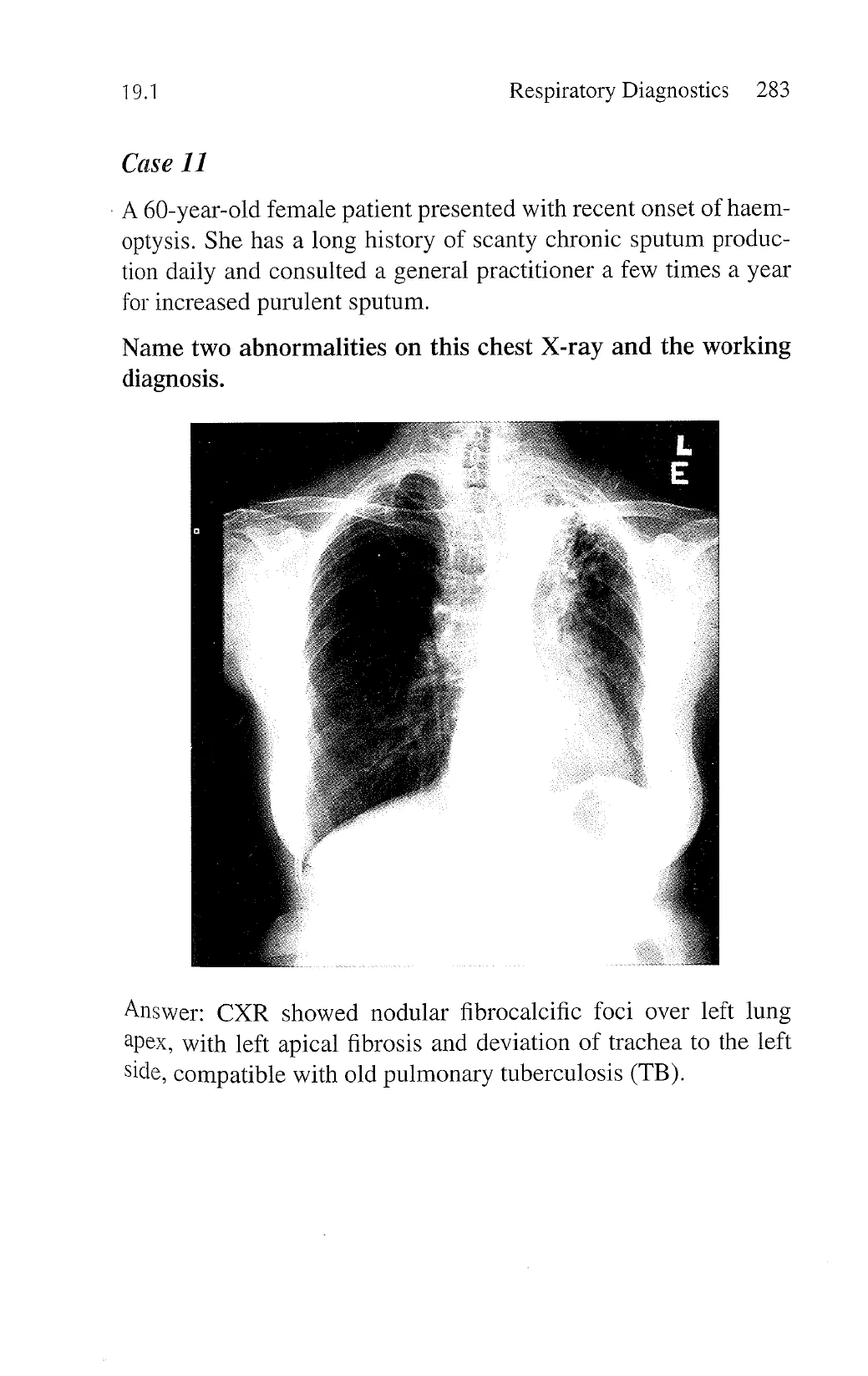
Name two abnormalities on this chest X-ray and the working
diagnosis.
Answer: CXR showed nodular fibrocalcific foci over left lung
apex, with left apical fibrosis and deviation of trachea to the left
side, compatible with old pulmonary tuberculosis (TB).
View original page (figures / layout)
Page 16
284 Problem-Based Medical Case Management 19.1
Name three investigations important for delineating the reason
for her haemoptysis.
1. Sputum for acid-fast bacilli and culture to look for reactivation
of TB.
2. Bronchoscopy to assess for airway lesion.
3. Computed tomography of the thorax—having both HRCT
and spiral CT would be useful to look for bronchiectasis and
further define parenchymal lesions as patient has new onset of
hemoptysis.
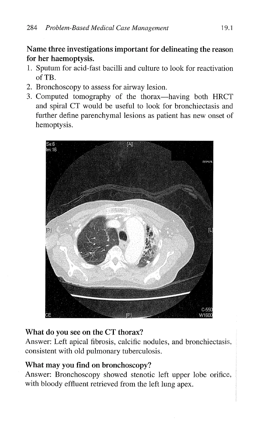
What do you see on the CT thorax?
Answer: Left apical fibrosis, calcific nodules, and bronchiectasis,
consistent with old pulmonary tuberculosis.
‘What may you find on bronchoscopy?
Answer: Bronchoscopy showed stenotic left upper lobe orifice,
with bloody effluent retrieved from the left lung apex.
View original page (figures / layout)
Page 17
19.2 Respiratory Diagnostics 285
19.2 Lung function tests
Case 1
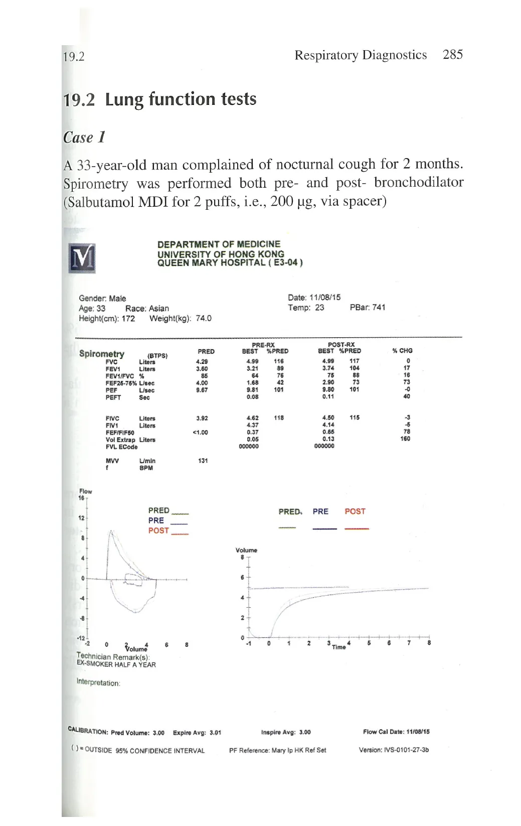
A 33-year-old man complained of nocturnal cough for 2 months.
Spirometry was performed both pre- and post- bronchodilator
(Salbutamol MDI for 2 puffs, i.e., 200 ug, via spacer)
DEPARTMENT OF MEDICINE
UNIVERSITY OF HONG KONG
QUEEN MARY HOSPITAL ( E3-04)
Gender: Male Date: 11/08/15
Age; 33 Race: Asian Temp: 23 PBar: 741
Height(cm): 172 Weight(kg): 74.0
PRERX POST-RX
Spirometry TP PRED BEST %PRED BEST %PRED % CHO
Fve Liters 4.29 498 16 499 "7 0
FEVY Liters 3.60 32 L 374 104 17
FEVIFVC % 8 o 7% 75 88 16
FEF26-78% Lisec. 4.00 1.68 42 290 73 kel
F Usec 8.67 9.851 101 9.80 101 0
PEFT Sec 0.08 011 @
Five Liters 382 462 118 450 18 -3
Fivi Liters 437 414 5
FEFIFIFSO <%0 037 088 78
Vol Extrap Liters 0.05 0.43 160
FVL ECode 000000 000000
M Umin 31
f BPM
16,
Al PRED __ PRED., PRE POST
2 PRE
T POST _—
8l
1 Volume
o 3
0 6+
« pul s
s
i 1 Fr s
o ?Inl “m'l 8 8 2 Time. 5 L] 7 8
Technician Remark(s)
EX-SMOKER HALF A YEAR
Interpretation:
CALIBRATION: Prod Volume: 3.00 Expire Avg: 301 Inspire Avg: 3.00 Flow Cal Date: 11108115
)= OUTSIDE 95% CONFIDENCE INTERVAL PF Reference: Mary Ip HK Ref Set. Version: IVS-0101-27-3b
View original page (figures / layout)
Page 18
286 Problem-Based Medical Case Management 19.2
Questions
1.
2.
Describe the spirometric abnormalities.
What is the diagnosis?
Answers
1.
2.
Mild airflow obstruction with significant reversibility after
bronchodilator (i.e., positive bronchodilator response).
Asthma
Description
1.
Reduced FEV, (at 89% of predicted when FVC was at 116%
of predicted) and reduced FEV,/FVC ratio (at 64% when the
predicted is 85%), compatible with airflow obstruction
. There was significant improvement in FEV, after bronchodila-
tor (530 ml and 17% increase), which was consistent with a':
diagnosis of asthma. '
Discussion
1.
The American Thoracic Society defines that a 12% or more
representing a 200 mL or more increase in FVC or FEV, in
the post-bronchodilator result above the pre-bronchodilator
state suggests a significant bronchodilator response in adults.
. The diagnosis of asthma is based upon the presence or history
of symptoms consistent with asthma (most commonly episodic .
cough, wheezing, or dyspnoea provoked by typical triggers).
The diagnosis of asthma is substantiated by the demonstration .
of variable expiratory airflow obstruction or a significant bron-
chodilator response in spirometry. However, a normal spirom
etry at stable state does not rule out asthma.
View original page (figures / layout)
Page 19
19.2 Respiratory Diagnostics 287
Case 2
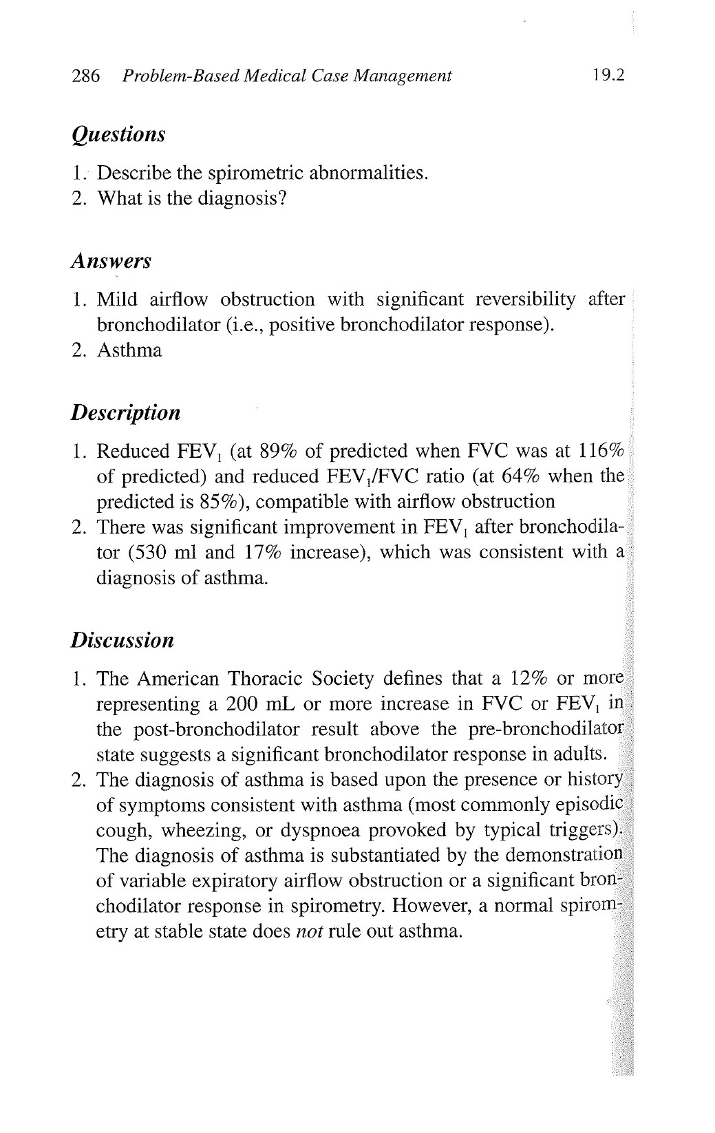
A 37-year-old woman complained of dry cough and progressive
shortness of breath on exertion for 3 months.
DEPARTMENT OF MEDICINE Page1of 1
UNIVERSITY OF HONG KONG
QUEEN MARYHOSPITAL (E3-04)
LUNG FUNCTION STUDY REPORT
Name: ABC, 1d: 01111
Gender; Female Date: 01/01/00
Age:37 Race: Asian Temp: 22 PBar: 743
Height(cm): 157 Weight(kg): 59.1 Physician:
Diagnosis: Technician:
Da Fl
Sty Ref Pre Pre Post Post Post fl
Mess % Ref Meas % Ref % Chg
FVC Liters 287 126 44
FEV1 Liters. 243 106 Bl
FEVIFVC % 85 B4 bl
FEF25-T5%L/sec 27 155 56
Livec 530 368 75
FEFTS% Usec 126 045 3
PEF Liec 697 43 &
FVC e 27 103 3
FIF50% Lisec 115
Lung Volume
e Liters 423 244
ve Liers 281 126 45
ic Liters 219 085 3
ERV Liters 110 033 30 1
RV Liters. 124 118 05 i
RUTLC % % 48 88 ]
Dfi;::gcaanw Hb: 106 grvdl. -, . - _
Q mUmmigmin 208 66 a2 -
DLAG mmmm: 208 713 35 N2 Vound
DLCOVA mlimHg/mink 516 363 70 &
Kroghs K 1/min 313
VA Liers a2 1 @
FICHs % 0300 ©
0.300
FECO & 0086 »
BHT Sec 1268
Ve e 106 2
L S S T s
¢ Voidme
ol — Volums DLCO (Volume & Gast% vs Time)
0 . 4 .
il 0.4
4 4 00
2 4]
0 e
[ EINAT T SR T
NON-s'fl'" Rumnk(s) T
LUNG VOLUME DONE BY N2 WASHOUT
| Intarprotation
lwmm.uarthKRefSet
View original page (figures / layout)
Page 20
288 Problem-Based Medical Case Management 19.2
Questions
1.
What are the abnormalities seen in this full lung function test?
2. Name one medical condition with these lung function test
findings.
Answers
1.
A restrictive pattern with reduced total lung capacity, functional
residual capacity, residual volume, and diffusion capacity from
carbon monoxide. Decreased KCO as well as DLCO suggests
parenchymal involvement. Flow rates (FEV, and FVC) are
reduced, and this is related to the decreased lung volumes. The
FEV /FVC ratio is normal.
. Interstitial lung disease (ILD).
Discussion
1.
In a patient of this sex (female) and age (37 years), one must
consider ILD including pulmonary fibrosis associated with con-
nective tissue diseases. ,
This lung function test pattern suggests diffuse parenchymal
lung disease, and the diagnosis of disease will depend on the
clinical features of the patient.
Patients are usually in hypoxaemic state, in type 1 respiratory -
failure. The resting arterial blood gas may be normal or showing .
low PO, (secondary to ventilation-perfusion mismatching) and -
respiratory alkalosis. .
View original page (figures / layout)
Page 21
19.3 Respiratory Diagnostics 289
19.3 Sleep case studies with polysomnograms
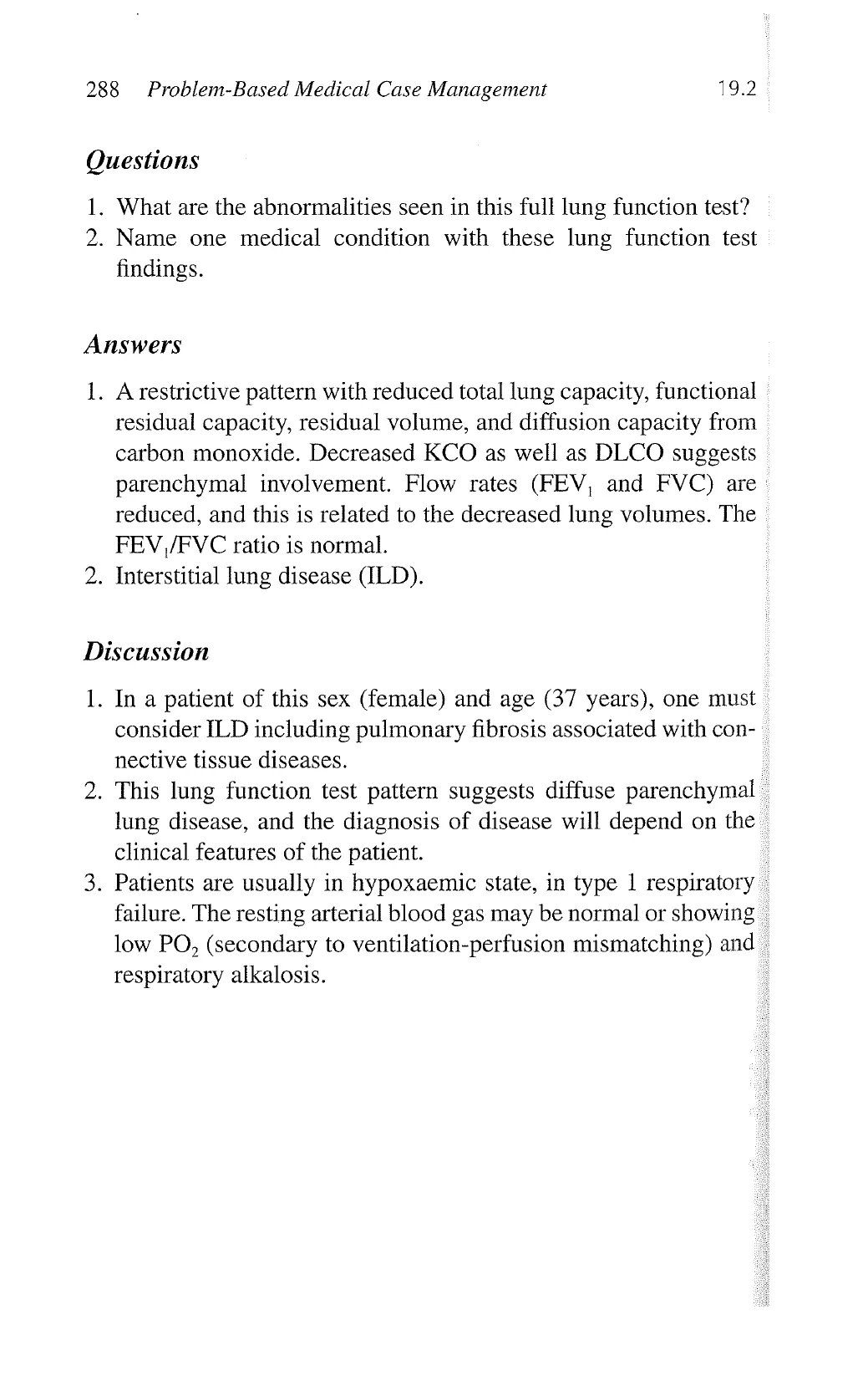
Case 1
A 65-year-old man presents with shortness of breath and bilateral
ankle oedema.
¢
sy
L
"
1
i
= i 1
3 i 3 iBER
i o = 9
H i
i 3
g o Ei
i .
3 - %
4 . 3
= P 3
= 3 R
= ~ 1 |
® 5
i il [ Fe
T =11 dk
4 L ks
5 » N
I
T
1
:
B
W
View original page (figures / layout)
Page 22
290 Problem-Based Medical Case Management 19.3
Question
What is shown in this epoch of polysomnographic tracing?
Answer
1. Cheyne-Stokes breathing with central sleep apnoea.
2. Cheyne-Stokes breathing is defined as a crescendo-decrescendo
pattern of breathing with central hypopnoea or apnoea at trough.
3. This may be associated with frequent arousals from sleep,
which occur at the highest point of ventilatory effort, rather
than at apnoea termination.
Discussion
The two most common causes of Cheyne-Stokes breathing are
congestive heart failure and neurological diseases.
Significant arterial oxygen desaturation is common in patients
with Cheyne-Stokes breathing during sleep, despite a normal
daytime oxygen saturation.
View original page (figures / layout)
Page 23
19.3 . Respiratory Diagnostics 291
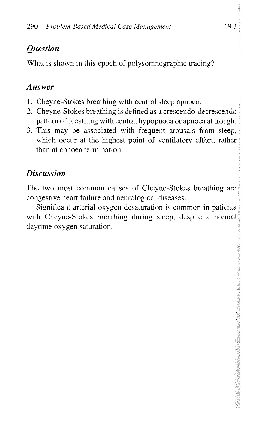
Case 2
This polysomnographic result belongs to a 50-year-old man who
kicks in sleep.
o] i
& o]l [nfnf o] ] ] 9]
[—
View original page (figures / layout)
Page 24
292 Problem-Based Medical Case Management 19.3
Question
What is the diagnosis?
Answer
Periodic limb movements (PLM) in sleep.
Description
There are repetitive right leg movements that occur at regular inter-
vals in this polysomnography (LEMG-R).
Some of these leg movements are associated with arousals.
Discussion
Periodic leg movements in sleep occur most commonly in sleep
stages 1 and 2, and less commonly during REM sleep.
Periodic leg movements can be idiopathic, or caused by end-
stage renal failure, pregnancy, iron deficiency, and certain drugs.
There may be an abnormality in iron transport into the central
nervous system or in use of iron in dopaminergic neuronal
transmission. ‘
PLM does not necessarily cause excessive daytime sleepiness,
and if so, it is called PLM disorder (PLMD). They should be dif-
ferentiated from restless leg syndrome (RLS), which is a clinical
diagnosis of daytime symptoms of “restless legs”.
View original page (figures / layout)
Page 25
293
Respiratory Diagnostics
ey
>
Case 3
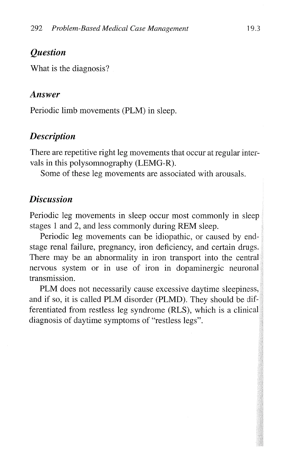
A 54-year-old man who presents with a 10-year history of exces-
sive daytime sleepiness.
ook % » (900 [z
5 - - - - - - - o - e
. . N . s el e
wmmmmmmmmmmmw(.mmwmwmmmmmmmm..e.wmmmmm
...:sssu.bnish.s. o
q 1
mamewm%wwmmmmmomammwmw“s
View original page (figures / layout)
Page 26
294 Problem-Based Medical Case Management 19.3
Question
What might this patient’s spouse notice during the recording
period?
Answer
¢ Episodes of cessation of breathing (apnoea).
¢ Interrupted snoring.
Description
Snoring is produced by vibration of the soft tissues of the upper
airway during sleep. In apnoea, there should be no airflow at
nose or mouth nor snoring due to complete cessation of airflow.
Snoring, however, may persist during partial collapse of upper
airway (in hypopnoea). She may notice persistent thwarted move-
ments of the thorax and abdomen.
Discussion
This tracing shows episodes of obstructive apnoea. Note that -
during an episode of obstructive apnoea, respiratory effort persists
(thoracic and abdominal movements), but there is no inspiratory
flow, and Sa0, drops progressively till a greater respiratory effort
(producing snoring) overcomes the obstruction temporarily and
results in rise of Sa0, again. The diagnosis of obstructive sleep
apnoea, however, cannot be made based on this tracing alone :
because at least 4 hours of sleep study is needed to document the -
total number of apnoea/hypopnoea during sleep. An apnoea/hypo-
pnoea index = 5 (total no. of apnoea/hypopnoea divided by total
sleep time in hours) is a commonly used definition for obstructive -
sleep apnoea. -
View original page (figures / layout)