Page 1
13
Respiratory Medicine—
Short Cases
David Chi-Leung Lam, James Chung-Man Ho,
and Mary Sau-Man Ip
Overview
The goals of clinical examination for the respiratory system, in the
format of short case examination, are:
1. To identify the abnormal physical signs.
2. To arrive at the pathophysiologic diagnosis underlying the
physical signs and suggest the most likely disease diagnosis or
differential diagnoses.
3. To propose, based on the working diagnosis, relevant clinical
investigations and briefly discuss salient management issues.
General examination
In the context of short case examination, this means periph-
eral examination for physical signs that are relevant to diseases
affecting the respiratory system.
Observe the overall patient status, facial expression, posture,
breathing pattern; also check for the presence of medical acces-
sories on the patient or at the bedside. For example:
1. Ventilators could be portable ones that patient may be using at
certain times of the day or at night and thus not dependent on
when you examine.
2. Oxygen supplementation vianasal cannula or face mask (Venturi
mask). Note the oxygen flow rate or oxygen concentration.
View original page (figures / layout)
Page 2
Respiratory Medicine 179
3, Medical drainage systems or intravenous drips.
4. Inhaler devices that patient could be using.
5. Sputum mug for collection of sputum.
6. Any bedside “alert signage” being put up.
Get an overall impression whether patient has cachexia/morbid
obesity, tachypnea or respiratory distress, such as excessive use of
accessory muscles of breathing or paradoxical breathing.
Look for pallor, central cyanosis, finger clubbing, ankle or leg
swelling, cervical and supraclavicular lymphadenopathy, skin
rash, facial/upper limb swelling with dilated veins over chest due
to superior vena cava obstruction syndrome, unilateral ptosis sug-
gesting possible Horner’s syndrome.
Do not restrict your focus on the signs being mentioned above
while ignoring gross abnormalities that the patient may show, e.g.,
rheumatoid hands which support pulmonary fibrosis.
Chest examination of the respiratory system
Important to note: As you approach the patient from either the front
or the back, do not miss the lung apices and the axillary regions,
where scars or chest drain sites could be hidden.
For the chest, compare the symmetry of findings on the two
sides, while taking note of presence of liver and heart.
* Inspection:
Look at the whole precordium including the base of the neck
(supraclavicular areas) and also the axillary region on both
sides. Observe, preferably from the end of the bed, any chest
wall deformity, thoracotomy scars, use of accessory muscles in
breathing, breathing pattern, and chest wall movement. (Note:
mid-sternotomy scars generally indicate previous cardiac
surgery or thymectomy).
Count the respiratory rate: though you may not be required
to do so for short case examination with very limited time—you
must have an observation of whether the patient is breathing too
rapidly or too slowly.
View original page (figures / layout)
Page 3
180 Problem-Based Medical Case Management Case 13.1
Palpation:
Assess chest wall expansion; the position of the trachea and the
apex beat; vocal fremitus (optional if vocal resonance will be
performed later; also suggest to put this test till the end if used
for confirmation).
Percussion:
To decide whether percussion note is resonant or dull
Auscultation:
Assess breath sounds (vesicular, bronchial; prolonged expira-
tion), detect added sounds (crackles or crepitations [fine or
coarse], thonchi or wheeze, pleural rubs), and assess vocal
resonance.
13.1 Bronchiectasis
Classical signs
.
Sputum container filled with purulent or blood stained sputum
at the bedside
Finger clubbing
Reduced chest wall movement
Normal percussion )
Bilateral or focal coarse inspiratory crackles that may disappear
or are altered in quality after coughing. (Note: crackles may
also be persistent). In some patients, rhonchi are also present.
Severe cases may be associated with features of cor pulmonale
and respiratory distress.
Very severe cases are often associated with cachexia.
Causes
Idiopathic (majority of diffuse bronchiectasis)
Post-tuberculosis
Post-pneumonic
Childhood measles (severe, complicated by pneumonia) or
pertussis
View original page (figures / layout)
Page 4
Case 13.1 Respiratory Medicine 181
Rheumatological disorders (rheumatoid arthritis, systemic
lupus erythematosis, systemic sclerosis, etc.)
Gastro-oesophageal reflux
Traction bronchiectasis from fibrotic lung diseases (such as
pulmonary fibrosis or radiation pneumonitis)
Kartagener’s syndrome
Hypogammaglobulinaemia
Allergic bronchopulmonary aspergillosis (ABPA)
Important differential diagnoses
For the physical finding of bilateral lung crackles, the major
dDx is pulmonary fibrosis. However, the crackles in bronchiec-
tasis are usually coarse and may occur through both inspiratory
and expiratory phases, while that in pulmonary fibrosis are fine
and mainly late inspiratory. Bronchiectasis may have rhonchi/
wheeze, and may show some hyperinflation while pulmonary
fibrosis results typically in reduced lung volumes (i.e., restric-
tive). Both can have finger clubbing.
Chronic obstructive pulmonary disease (COPD) with heavy
component of chronic bronchitis may present with similar
symptoms of cough and sputum in a smoker. Studies have actu-
ally shown a high prevalence of bronchiectasis among patients
diagnosed to have COPD. COPD usually have more hyperinfla-
tion and less coarse crackles.
Asthma presents with dyspnoea, cough, sputum production,
and recurrent exacerbations. Asthma affects up to 10% of
patients with bronchiectasis and therefore asthmatic patients
with copious sputum production should also undergo high
resolution computed tomography (HRCT) examination of the
chest. Physical examination may show both wheeze/rhonchi in
addition to crackles in patients with only bronchiectasis or with
both bronchiectasis and asthma.
Post-tuberculosis lung destruction with a mixture of bronchiec-
tasis, fibrosis, and cavitations. Usually causes focal/unilateral .
bronchiectasis.
View original page (figures / layout)
Page 5
182 Problem-Based Medical Case Management Case 13.1
Tnvestigations:
e CXR and HRCT thorax for definitive diagnosis
» Spirometry (may be obstructive or mixed obstructive-restrictive)
e Sputum microscopy, Gram’s stain and bacterial and fungal
culture, smear and culture for acid-fast bacilli
« Sa0, (on room air) or arterial blood gases determination
« X-ray sinuses
¢ Other tests as necessary (e.g., aspergillus serology)
Discussion
« Aectiology of bronchiectasis:
— In Hong Kong and the West, bronchiectasis is largely an
idiopathic disease. However, TB, pneumonia, and immuno-
deficiency also account for a significant proportion.
— Cystic fibrosis is extremely rare in Chinese.
«+ Differential diagnoses and key distinguishing investigations
(see Chapter 14)
+ Management in acute exacerbations or in severe cases
Pitfalls and tips
« Failure to recognize nasal tone of chronic nasal obstruction
(or other signs of chronic sinusitis)
« Failure to detect (i) mediastinal shift in severe bronchiectasis;
resulting from associated lobar collapse, or (ii) Situs inversus in
particular dextrocardia in Kartagener’s syndrome
+ Omitting examination of bedside sputum pot
Patients with idiopathic pulmonary fibrosis may have “traction
bronchiectasis” resulting in mixed features of pulmonary fibro-
sis and bronchiectasis.
View original page (figures / layout)
Page 6
Case 13.2 Respiratory Medicine 183
13.2 Chronic obstructive pulmonary disease
(COPD)
Classical signs
Central cyanosis (may be masked by use of supplemental
oxygen)
Bilateral pitting ankle oedema and other features of right heart
failure
Tachypnea
Reduced distance between suprasternal notch and cricoid carti-
lage (normal distance depends on individual body build, about
2-3 fingers’ breadth) and increased anteroposterior diameter of
the chest (barrel chest) with reduced chest expansion bilater-
ally, due to hyperinflation of the lungs
Normal or hyper-resonant percussion (obliteration of cardiac
and liver dullness)
Decreased breath sounds (with prolonged expiratory phase, end
expiratory rhonchi or inspiratory crackles depending on the
severity of the airflow obstruction and the amount of sputum
present)
Normal to decreased vocal resonance
Risk factors
Cigarette smoking: the single most important risk factor
Heavy exposure to indoor air pollution, e.g., use of biomass
fuels in some rural areas
Alpha-1 antitrypsin deficiency: rare in Chinese; this condition
should be suspected in young persons (< 50 years old) with
COPD who never smoked or were light smokers.
Occupational exposures to dusts or fumes
Important differential diagnoses
* Chronic asthma: both chronic asthma and COPD show airflow
obstruction but the former exhibits greater variation in symptoms
View original page (figures / layout)
Page 7
184 Problem-Based Medical Case Management Case 13.2
and reversibility of airflow obstruction on spirometry. Asthma
usually affects non-smokers with a history of wheezing since
childhood.
« A group of patients (usually smokers) may have overlapping
features of asthma and COPD (ACOS).
¢ Bronchiectasis: both bronchiectasis and COPD may present
with chronic cough and sputum (chronic bronchitis component
of COPD), and have lung crackles and rhonchi on physical
examination. However, patients with bronchiectasis usually
are non-smokers, show symptoms dating back to childhood or
youth, and produce copious purulent sputum. Bronchiectasis
and COPD may also coexist.
Important investigations
+ Chest X-ray: typical COPD features include hyperinflation and
bullae; typical complications include infections, pneumotho-
rax, cardiomegaly (due to cor pulmonale), prominent pulmo-
nary trunks (pulmonary hypertension), and lesions suspicious
of lung cancer (another complication of chronic smoking, the
main cause of COPD).
« Pulse oximetry (assesses Sa0,) or arterial blood gases (assesses
pH, PaO, and PaCO,): to confirm/explore severity and type
(1 or 2) of respiratory failure
« Lung function tests:
— Spirometry with bronchodilator reversibility test to confirm
and assess severity of airflow obstruction (typically FEV1/
FVC < 70%, FEV1 < 80% of predicted)
— Lung volume measurement to assess severity of hyperinfla-
tion (RV and RV/TLC)
- Diffusing capacity of carbon monoxide (DLco) to assess
alveolar destruction due to emphysema
» Complete blood count: raised haematocrit indicates erythrocy-
tosis secondary to chronic hypoxaemia; high leucocyte count
may indicate acute bacterial infection and exacerbation or use
of systemic steroids.
View original page (figures / layout)
Page 8
Case 13.3 Respiratory Medicine 185
Sputum examination: for patients with infective exacerbations.
Note: Sputum culture is not routinely required, as the com-
monest isolates are known to be Streptococcus pneumoniae,
Haemophilus influenzae and Moraxella catarrhalis. Antibiotics
to cover these pathogens are prescribed whenever there is a
clinical diagnosis of an infective exacerbation.
Nasopharyngeal aspirate for virus.
ECG and echocardiogram: to assess right heart strain and cor
pulmonale.
Pitfalls and tips
Look out for sputum container and contents, and presence of
inhaler devices or non-invasive ventilators by the bedside.
Note whether patient is receiving oxygen usually via nasal
cannula (most are), and the rate of supplemental oxygen.
Note whether patient is in respiratory distress (rapid and
shallow breathing, using accessory respiratory muscles at the
neck, intercostal retraction) or drowsy (sign of CO, retention or
exhaustion).
Note “pursed lip breathing” if present.
Note “barrel chest” if present.
In patients with severe airflow obstruction (including in
asthma), the expiratory phase is prolonged. Expiratory rhonchi/
wheeze may not be apparent unless the patient is requested to
carry out a forced full expiration.
Note physical signs of cor pulmonale.
13.3 Collapsed lung
Classical signs
Tachypnea, especially in case of major collapse
Decreased chest movement on affected side
Tracheal deviation and shifting of apex beat to ipsilateral side.
Tracheal deviation occurs in whole lung collapse or upper lobe
View original page (figures / layout)
Page 9
186 Problem-Based Medical Case Management Case 13.3
collapse, but not obvious in lower lobe collapse; while apex
beat is affected more by lower lobe or whole lung collapse.
Dull to percussion (not “stony”) over affected area
Decreased breath sounds over affected area
Causes
Endobronchial obstruction or extrinsic compression by neo-
plasms, e.g., lung cancer, rarely bronchial adenoma.
Foreign body aspiration
Infections, e.g., endobronchial tuberculosis (may present as
sequelae of previous tuberculosis)
Impacted, thick tenacious sputum, usually in weak and ill
patients
Important investigations
.
Chest X-ray: confirms lung collapse radiologically. Then look
for mass lesion compressing airway; some foreign bodies are
radio-opaque.
CT thorax can reveal collapse and mass lesion compressing
airway and associated mediastinal lymphadenopathy.
Sputum: cytology for malignant cells and smear and culture for
acid-fast bacilli
Bronchoscopy: to localize obstruction and obtain specimens for
diagnosis
Discussion
Treatment depends on the aetiology. Lung cancer causing bron-
chial obstruction: requires surgery (if resectable), radiotherapy
and/or medical therapy, bronchoscopic stenting; foreign body
requires bronchoscopic removal; active endobronchial tuber-
culosis mandates prompt anti-tuberculosis chemotherapy to
minimize fibrosis and airway stenosis (use of systemic corti-
costeroids is controversial).
View original page (figures / layout)
Page 10
Case 13.4 Respiratory Medicine 187
Pitfalls and tips
Overlooking cervical lymphadenopathy, especially for sus-
pected malignancy
Tracheal deviation and shifted cardiac apex related to upper and
lower lung collapse, respectively
Pleural effusion and lower lobe collapse have similar physical
signs except for the degree of dullness, and pleural effusion
may be associated with underlying lung collapse giving rise to
stony dullness. The directions of mediastinal shift, if present,
will be ipsilateral to the side of disease in collapse but to the
contralateral side if due to pleural effusion.
Signs of lower lobe collapse and lobectomy may be similar,
though lobectomy should be accompanied by thoracotomy scar
or a few small scars of VATS.
In consolidation-collapse, features of consolidation such as
localized crackles or even bronchial breathing may be present.
Endobronchial tuberculosis (alone or as part of pulmonary
parenchymal tuberculosis) is an important benign cause of
obstruction, still not uncommon in less developed places. It may
be mistaken for tumour on gross bronchoscopic examination.
Some patients present with lung collapse as late sequelae of
previous tuberculosis.
13.4 Pleural effusion
Classical signs
Tachypnea
Decreased chest movement or expansion on the affected side
Tracheal deviation to the contralateral side (if massive)
Stony dull percussion note on the affected side
Decreased/absence of breath sounds on the affected side; there
may be bronchial breath sounds at the upper margin of effusion
(egophony)
Decreased vocal resonance
View original page (figures / layout)
Page 11
188 Problem-Based Medical Case Management Case 13.4
Common predisposing scenarios/causes
.
Young person with pleurisy due to tuberculosis or lupus
Patients with diabetes or on systemic corticosteroids are liable
to effusions from bacterial, tuberculous or fungal pleurisy
Elderly male chronic smoker (malignant effusion, tuberculous
pleurisy)
Chronic smoker and alcoholic (malignant effusion, tuberculous
pleurisy, suppurative aspiration pneumonia with empyema)
Occupational exposure to asbestos (mesothelioma)
If bilateral, consider: heart failure and kidney disease (transu-
dates). Hepatic hydrothorax (cirrhosis), though transudative,
is mostly right-sided.
Important investigations
Chest X-ray (may include decubitus view for small effusion):
to confirm unilateral effusion and detect parenchymal shadows
suspicious of tuberculosis or malignancy, mediastinal lymphad-
enopathy; rib erosions suggest a malignant process.
Ultrasound is useful for further delineation of pleural effusion
to guide thoracocentesis.
Sputum examination for: cytology, smear and culture for bacte-
ria, acid-fast bacilli and fungi as indicated
Diagnostic thoracocentesis for:
— Inspection of the fluid is important. Turbid fluid should raise
concerns of empyema, chylothorax or pseudochylothorax. -
Bloody effusion may be a haemorrhagic effusion from
malignancy or due to a haemothorax. Presence of food parti-
cles indicates oesophageal rupture. ,
— Biochemistry, including serum/fluid protein and LDH to
ascertain exudative or transudative nature
— Fluid white cell count and differential (neutrophil predomi-
nance — acute inflammation, e.g., bacterial infection; lym-
phocyte predominance — tuberculous or vasculitic pleurisy
View original page (figures / layout)
Page 12
Case 13.4 Respiratory Medicine 189
such as in lupus). Malignant effusions can be lymphocytic or
neutrophilic.
— Culture for bacteria/mycobacteria/fungi (note that smear of
pleural fluid for acid-fast bacilli is negative as a rule, even
in tuberculous pleurisy (except in HIV-infected subjects).
TB-PCR is not recommended due to interference from
inhibitors in the fluid with poor sensitivity and specificity).
— Cytology for malignant cells
— Adenosine deaminase level (higher than reference range)
suggests tuberculous effusions although ADA can be ele-
vated in most effusions of bacterial infection, and some of
those from rheumatological causes and cancers.
Note: Such aspiration procedures are often followed by a
pleural biopsy if effusion is exudative (to look for granuloma-
tous tuberculosis or a malignant pathology). In difficult diag-
nostic cases, pleuroscopy/VATS may be needed.
* CT thorax for better delineation of lung parenchyma and effu-
sion as indicated. If clinically indicated, CT pulmonary angio-
gram may be needed as some patients with pulmonary emboli
will have a (usually small) pleural effusion.
* Flexible bronchoscopy if indicated
* Search for autoimmune disease markers as indicated.
Pitfalls and tips
* Presence of an aspiration needle mark (usually posteriorly) pro-
vides an additional clue to the presence of an effusion.
Mediastinal shift (tracheal deviation) may not be present with
small to moderate sized pleural effusions. Mediastinal shift is
also modified by presence of both lung collapse and pleural
effusion which may occur together.
In very weak, bed-bound patients, percussion dullness is
detected more in the lateral (dependent) parts of the chest.
View original page (figures / layout)
Page 13
190 Problem-Based Medical Case Management Case 13.5
13.5 Upper lobe/apical fibrosis
Classical signs
« Post-radiotherapy skin changes around the neck or upper chest
area
¢ Flattened chest on the affected side (increased supraclavicular
fossa hollowing)
e Decreased chest movement on the affected side (area)
¢ Tracheal deviation to the affected side
« Dull percussion note, particularly over the clavicle
» Decreased breath sounds on the affected side with localized fine
crackles. Occasionally bronchial breath sounds are present.
Common causes
¢ Old pulmonary tuberculosis
« Ankylosing spondylitis
¢ Post-radiation pulmonary fibrosis (e.g., for nasopharyngeal car-
cinoma or lung cancer)
Important investigations
* Chest X-ray (may include apico-lordotic view): confirms fibro-
sis in lung apex, calcified apical foci suggest old pulmonary
tuberculosis, spine may have features of ankylosing spondylitis.
» Computed tomography (CT) of thorax: delineates parenchymal
lung lesions better [
« Sputum smear and culture for acid-fast bacilli: assess activity of
pulmonary tuberculosis
Pitfalls and tips
¢ Chest wall expansion may be only mildly diminished, and signs -
rather subtle if fibrosis only involves apical area. The only
detected sign may be percussion dullness over the clavicle with
or without deviation of trachea to affected side. ‘
View original page (figures / layout)
Page 14
Examination case Respiratory Medicine 191
«+ Do not forget percussion over the clavicles.
+ PFeatures of upper lobe collapse or collapse-consolidation due
to endobronchial obstruction, e.g., by tumour, may give rise
to similar features of tracheal deviation, dull percussion, and
decreased breath sounds (or bronchial breath sounds when
extensive consolidation is present).
+ Uncommonly, similar signs are due to Pancoast tumour.
Associated Horner’s syndrome may provide the hint.
Discussion
+ Previous pulmonary tuberculosis is a common cause of apical
or upper lobe fibrosis in Asians.
* There is a need to ascertain any previous investigation and man-
agement for pulmonary tuberculosis, including the treatment
regime and whether directly observed therapy or “DOT” was
adopted (as practised in Hong Kong).
Examination case scenarios
i. Examine this lady’s lungs. She complained of
progressive shortness of breath for a year.
Important signs » Tachypneic and using oxygen at
2 litres/minute.
* Mild finger clubbing.
* Palpation and percussion of chest
normal.
» Fine end inspiratory crackles in lower
zones bilaterally in the posterior and
lateral aspects.
Diagnosis Idiopathic pulmonary fibrosis
View original page (figures / layout)
Page 15
192 Problem-Based Medical Case Management
Examination case
Questions
1. What other diseases commonly give
rise to clubbing together with inspiratory
crackles?
¢ Bronchiectasis.
¢ If localized coarse crackles—may also
be obstructive pneumonia due to lung
cancer.
2. What other features would you look
for systemically in case of pulmonary
fibrosis?
¢ Features of autoimmune disease such as
rheumatoid arthritis.
3. What investigation would be most
useful to confirm the diagnosis?
* High resolution CT thorax.
4. What would you see on lung function
test?
* Restrictive pattern with decrease
diffusing capacity for carbon monoxide.
Pitfalls and tips
Remember to auscultate for added lung
sounds at the lateral aspects.
» Traction bronchiectasis may be present
in advanced pulmonary fibrosis.
« Typically, features of lung restriction are
present in advanced pulmonary fibrosis.
« Look for features of autoimmune disease
and also occupational exposure/drug
history.
» Coarse crackles and production of
purulent sputum suggest bronchiectasis
as dDx or added component.
View original page (figures / layout)
Page 16
Examination case
Respiratory Medicine 193
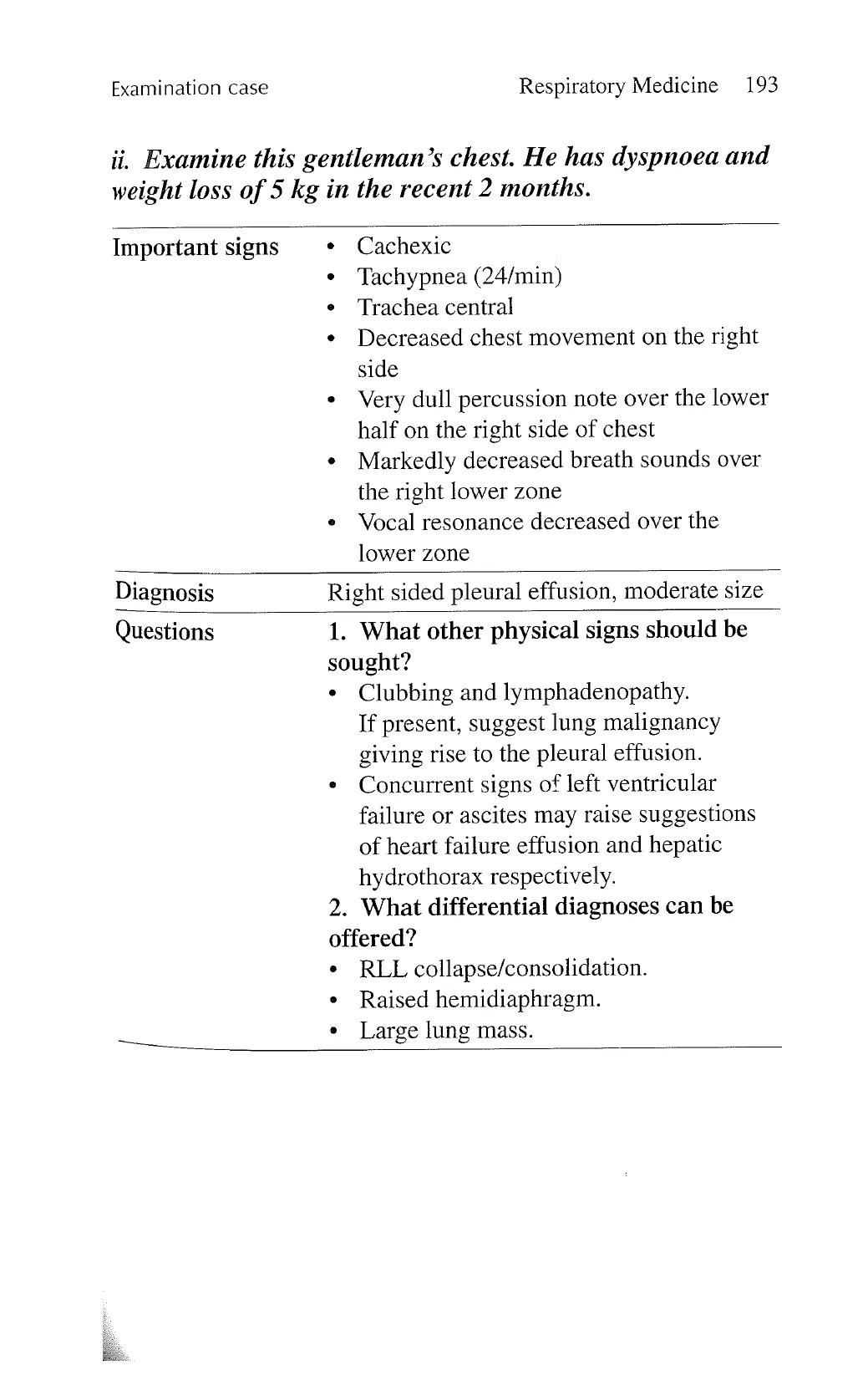
ii. Examine this gentleman’s chest. He has dyspnoea and
weight loss of 5 kg in the recent 2 months.
Important signs .
.
Cachexic
Tachypnea (24/min)
Trachea central
Decreased chest movement on the right
side
Very dull percussion note over the lower
half on the right side of chest
Markedly decreased breath sounds over
the right lower zone
Vocal resonance decreased over the
lower zone
Diagnosis Right sided pleural effusion, moderate size
Questions 1.
What other physical signs should be
sought?
2
Clubbing and lymphadenopathy.
If present, suggest lung malignancy
giving rise to the pleural effusion.
Concurrent signs of left ventricular
failure or ascites may raise suggestions
of heart failure effusion and hepatic
hydrothorax respectively.
What differential diagnoses can be
offered?
RLL collapse/consolidation.
Raised hemidiaphragm.
Large lung mass.
View original page (figures / layout)
Page 17
194 Problem-Based Medical Case Management Examination case
iii. Examine this woman’s chest. She had no known
respiratory disease but was admitted two days ago with
hypercapnic respiratory failure.
Important signs ~ + Obese (BMI = 35 kg/m”)
* Waist circumference: 103cm
» Central cyanosis
< Bilateral pitting ankle oedema
« Symmetrically decreased chest
expansion, but no wheeze or crackles
¢ Blood pressure 150/100 mmHg
¢ Left parasternal heave
¢ Increased pulmonic second sound
Diagnosis Obesity hypoventilation syndrome (OHS)
Questions 1. What are the likely underlying
respiratory abnormalities?
* Obesity hypoventilation syndrome
(presence of hypercapnia and cor
pulmonale).
« Obstructive sleep apnoea often
co-existent in OHS.
2. What is the treatment of choice in the
acute admission and in the long term?
» Non-invasive ventilation would control
the hypercapnic respiratory failure. Long
term—use either bi-level or continuous
positive airway pressure (CPAP) during
sleep.
» Supplemental oxygen is often needed for
hypoxemia in cases of OHS.
¢ Weight reduction.
» Treat right heart failure.
View original page (figures / layout)