Page 1
14
Respiratory Medicine—
Long Cases
James Chung-Man Ho, Christopher Kim-Ming Hui,
David Chi-Leung Lam, Kenneth Wah-Tak Tsang, Wah-Kit Lam,
and Mary Sau-Man Ip
14.1 Persistent cough with dyspnoea (asthma)
History
A 25-year-old waitress working in a restaurant presented with
persistent cough and occasional shortness of breath for several
months. Chest radiograph showed no lung lesion. Spirometry
revealed an obstructive pattern of deficits. She was a non-smoker.
She was diagnosed to have asthma, and a prescription of moderate
doses of inhaled corticosteroids and a beta-2 agonist was given for
symptomatic relief. At clinic follow-up 2 months later, she com-
plained of persistent symptoms.
Physical examination
No abnormality was detected at the time of clinic consultation.
No wheeze or prolonged expiratory phase was evident even on
forced expiration.
Questions
L. What factors should be explored to optimize treatment of
her asthma?
* Compliance to medications should be checked, particularly to
her preventive drugs (inhaled corticosteroids). Patients do not
View original page (figures / layout)
Page 2
196 Problem-Based Medical Case Management Case 14.1
comply for various reasons, including: poor understanding of
drug actions, fear of side effects, inconvenience.
The technique for using her inhaler(s).
Possible aggravating factors/triggers:
— New or unidentified allergens (e.g., new curtains/carpets,
pets at home, food additives).
— Occupational exposure.
— Cigarette smoking.
— Indoor/outdoor air pollutants (her job in a restaurant may
have exposed her to considerable passive smoking).
— Drugs, e.g., aspirin and other NSAIDs, beta-blocker.
— Emotional triggers.
Upper airway problems: rhinitis, sinusitis.
Consideration for coexisting diseases: such as gastro-oesopha-
geal reflux and/or thyrotoxicosis.
2. What other treatment modifications should be considered
at this stage?
Addition of inhaled long-acting beta-2 agonist to the regime.
This may be given in a separate inhaler, or in an ICS/LABA
combination inhaler, which has the advantage of avoiding
exclusive use of LABA without ICS by the patient.
Other additional treatment options include: sustained-release
theophyllines; long-acting oral beta-2 agonist; increasing the
dose of inhaled corticosteroids; or using leukotriene antagonist.
Novel therapies in the pipeline include anticytokine therapies
and bronchial thermoplasty.
Note: Refer to GINA guidelines for further stepwise treatment
titrations.
Pitfalls and tips
Some conditions can mimic asthma. A patient with “asthma
refractory to usual treatment” should have the diagnosis care-
fully reviewed and reassessed in a specialist clinic. Conditions -
that may be mistaken for asthma include:
— Upper airway obstruction: laryngeal stenosis, tumours ofr
other lesions affecting the upper airway or major bronchi,
View original page (figures / layout)
Page 3
Case 14.2 Respiratory Medicine 197
or vocal cord dysfunction. Breathing difficulty is typically
inspiratory.
— Lower airway: obstructive airway diseases such as bronchi-
ectasis, COPD, bronchiolitis obliterans, diffuse panbronchi-
olitis (DPB).
— Conditions with asthma as only one of the components:
allergic bronchopulmonary aspergillosis, eosinophilic gran-
ulomatous pulmonary angiitis (EGPA) previously known
as Churg-Strauss Syndrome, carcinoid (asthma component
may or may not be difficult to manage, but other manifesta-
tions of the disorder may dominate).
- Left heart failure.
+ A few individuals have very troublesome asthma that is not
fully amenable to control despite optimization of conventional
treatment. Such patients may be controlled with long-term
systemic steroids (steroid-dependent asthma), and despite such
treatment others still remain poorly controlled (steroid-resistant
asthma).
* When NOT having an acute attack, some patients have very
subtle findings of airflow obstruction. In which case, rhonchi
or a prolonged expiratory phase may only be detected after a
forced expiration.
14.2 Recurrent haemoptysis (bronchiectasis)
History
A 56-year-old woman with a 20-year history of recurrent episodes
Qf lower respiratory tract infection and chronic sputum produc-
tion was admitted following a 2-week period of increasing cough,
sputum production, and haemoptysis. Relevant history included
Chfonic rhino-sinusitis and recurrent exacerbations of haemoptysis
With dyspnoea complicating upper respiratory tract viral infec-
tions. She had no organ specific autoimmune disorders (such as
rl‘leumatoid arthritis or systemic lupus erythematosus), or any
;' history of pertussis, measles, tuberculosis or fibrotic lung disease.
View original page (figures / layout)
Page 4
198 Problem-Based Medical Case Management Case 14.2
Physical examination
There were coarse inspiratory crackles over the lower zones of
both lungs. There was a pot of purulent sputum at the bedside. The
patient had a “chesty” cough, mild finger clubbing but no cervical
lymphadenopathy, cyanosis or signs of heart failure. Precordial
examination revealed a loud pulmonic second heart sound, an
undisplaced cardiac apex and absence of parasternal heave or heart
murmur.
Questions
1. How would you investigate this patient?
» Confirm the presence of bronchiectasis and its severity by
high-resolution CT (HRCT) of the thorax. Its severity can be
gauged by lung function tests as well as clinical features such
as exacerbation frequency and 24-hour sputum volume. The
aetiology may be further evaluated by determining immuno-
globulin levels, studying gastro-oesophageal reflux, and col-
lecting sputum for acid-fast bacilli smear and culture. Further
investigations can be undertaken as indicated.
2. What do the chest X-ray and HRCT scan show?
¢ The plain chest X-rays may show “tram-lines” due to thicken-
ing of bronchial walls in cylindrical bronchiectasis. This radio-
logic feature is not highly specific and only has a sensitivity of
~47% compared to bronchography (an investigation now
superseded by CT imaging). There may also be hyperinflation.
In severe cystic bronchiectasis, cystic dilatations =+ fluid levels
due to copious secretions in the bronchi are characteristically -
seen. In Kartagener’s syndrome, there is dextrocardia = situs
inversus. '
In bronchiectasis, HRCT may reveal that affected airway inter- -
nal lumens are larger than their accompanying arteries, giving -
rise to a “signet-ring” appearance. Other features include: lack
of normal bronchial tapering, presence of peripheral airways
within 1 cm of the costal pleura, air-fluid levels in dilated
bronchi, and linear clusters of cysts (refer to Chapter 19 for
relevant imaging examples). ‘
View original page (figures / layout)
Page 5
Case 14.2 Respiratory Medicine 199
3. What is bronchiectasis?
+ Bronchiectasis is a syndrome of chronic cough and daily viscid
sputum production associated with airway dilatation and bron-
chial wall thickening. Multiple conditions are associated with
the development of bronchiectasis, but all require an infectious
insult and usually also impairment of drainage, airway obstruc-
tion, and/or a defect in host defence.
+ Of the broad spectrum of causes of non-cystic fibrosis bron-
chiectasis, only a few respond to direct treatment (e.g., certain
immunodeficiencies, non-tuberculous mycobacterial infection,
allergic bronchopulmonary aspergillosis). Instead, treatment
of bronchiectasis is aimed at controlling infection, reducing
inflammation, and improving sputum clearance. Surgical extir-
pation of affected areas may only be useful in a small number
of carefully selected patients.
. What is the lung function test pattern in bronchiectasis?
Obstructive, or obstructive-restrictive.
. What are the important sputum pathogens?
Haemophilus influenzae in early disease, and Pseudomonas
aeruginosa in late disease.
6. Suggested antibiotics useful for exacerbations of
bronchiectasis.
* For early/mild cases, amoxicillin and clavulanic acid
(Augmentin®) may be used against Haemophilus influenzae;
7-10 days of therapy are adequate. For late/more severe cases
(with frequent exacerbation and copious sputum production)
adequately high doses of levofloxacin or other IV antibiot-
ics directed against Pseudomonas aeruginosa are preferred
and given for 10-14 days (or longer) until sputum production
returns to baseline volumes.
Attention must be paid to pathogen resistance as this emerges
almost invariably over the time-course of treatment. Aside
from traditional modes of resistance and wild-type selection,
organisms such as Pseudomonas aeruginosa are also able to
produce a polysaccharide biofilm which resists penetration
by many antibiotics to which it would otherwise be sensitive.
Once formed, the biofilm matrix represents a critical interface
between the bacterium and the host or its environment.
LY LN
View original page (figures / layout)
Page 6
200 Problem-Based Medical Case Management Case 14.2
7. What is the long-term management?
+ Suitable antibiotic therapy should be promptly instituted for
each exacerbation, and diligent sputum clearance at home with
chest physiotherapy and postural drainage should be done as
retained mucus is harmful.
» Treatable causes or associated conditions such as host immuno-
deficiency, gastro-oesophageal reflux, asthma should be looked
for and tackled appropriately.
* Beta-2 agonists have been clinically utilized for patients with
obstructive defect on lung function testing, and have a theoreti-
cal advantage of enhancing ciliary clearance.
* Inhaled corticosteroid has been shown to reduce sputum inflam-
matory indices and may be tried in individual cases. It should
not be routinely continued if no clinical improvement is seen.
» The disease is largely irreversible. Severe bronchiectasis may
lead to premature death, and should be managed in specialist
clinics. To prevent further damage and to enhance quality of life,
anti-inflammatory treatment has been used. Macrolides (eryth-
romycin, clarithromycin or azithromycin) have been shown
in randomized controlled trials to reduce sputum volume and
to reduce exacerbations. Prolonged QTc and hearing impair- -
ment are potential side effects that need to be monitored.
* In very severe cases constantly producing copious sputum
with frequent exacerbations, a pre-emptive regime of periodic
intravenous antibiotic regime or long-term nebulised antibiotics
may be considered. .
* For severe advanced disease, suitable cases should be referred
for consideration of lung transplantation.
8. How do you manage haemoptysis in bronchiectasis?
* Bed rest, with hospitalization for severe haemoptysis. .
¢ Antibiotic courses are given (as detailed above) until sputumf‘i
volumes or coughing returns to baseline values and/or fever -
settles. -
* Use of thrombotic agents (such as tranexamic acid [Transamin®])
are not recommended routinely, as tenacious blood clots could
obstruct airways (leading to lung collapse, further infection,
and respiratory failure). Besides, by inducing thrombosis in
View original page (figures / layout)
Page 7
Case 14.3 Respiratory Medicine 201
blood vessels, they could give rise to stroke, myocardial infarc-
tion or peripheral vascular obstruction.
« Bronchial artery embolization is done for prolonged and severe,
or for massive life-threatening hemoptysis.
+ Very occasionally, surgical resection of the diseased actively
bleeding lung segment may be required, though this is usually
not feasible in view of diffuse disease involvement.
Pitfalls and tips
+ It may be misdiagnosed as COPD, asthma or both.
+ 10% of bronchiectasis cases may have some degree of asthma,
in whom anti-asthma medications have good treatment effect.
+ Haemoptysis ensuing in a patient with bronchiectasis should
not be ignored, as it indicates exacerbation of bronchiectasis,
and it might also indicate bronchial neoplasm or tuberculosis,
especially in new onset of haemoptysis.
+ Treatment with antibiotics for exacerbations alone may not be
adequate to control disease activity; and more intensive regular
treatment as outlined above is needed.
* Learn about the different classes of anti-pseudomonal
antibiotics.
* Development of antibiotic resistant strains and biofilm forma-
tion through wild-type selection and the use of antibiotics is a
significant problem for long-term treatment efficacy.
14.3 Chronic cough and weight loss (carcinoma
of lung)
History
A 50-year-old chronic smoker presented with 1-month history
9f persistent cough and blood-stained sputum. He complained of
Increasing shortness of breath, and a sharp pain in his right chest
“fhen he breathed. There was no hoarseness of voice, headache,
limb weakness or bone pain. He noted a weight loss of 8 kg in the
View original page (figures / layout)
Page 8
202 Problem-Based Medical Case Management Case 14.3
last 3 months. Chest X-ray showed an opacity at the right middle
zone of the lung field. It was later confirmed to be carcinoma of the
lung. The patient was admitted for further investigations.
Physical examination
There was a palpable 2 cm lymph node in the right cervical region
and clubbing of the fingers. Chest examination showed right mid
zone dullness on percussion with reduced air entry. There was no
hepatomegaly or focal neurological abnormality.
Questions
1. What differential diagnoses can be offered?
e Carcinoma of right lung, metastatic cancer to the lung, pneumo-
nia, and pulmonary tuberculosis.
2. What are the appropriate investigations?
* Sputum for cytology (presence of malignant cells, small cell
versus non-small cell carcinoma) and sputum smear and cul-
tures for acid-fast bacilli (tuberculosis) should be performed.
¢ A biochemical screen for deranged liver function (sugges-
tive of liver metastases), raised alkaline phosphatase (sug-
gestive of bone metastases) with/without hypercalcaemia
(can occur from PTH-like peptide release from lung carcino-
mas), hyponatremia (syndrome of inappropriate anti-diuretic
hormone).
¢ Contrast computed tomography scan of the thorax (including
upper abdomen) for baseline staging of lung cancer. Computed
tomography scan or MRI of the brain in case of clinical suspi-
cion of brain metastases.
* Incase of bone pain: look out for raised serum calcium and alka-
line phosphatase levels, a bone scan should also be considered. -
* Positron emission tomography (PET scan) is a useful whole-
body metabolic imaging procedure, which may be useful for
staging and detecting unsuspected metastasis, thus avoiding
unnecessary thoracotomy. .
View original page (figures / layout)
Page 9
Case 14.3 Respiratory Medicine 203
« Diagnostic tests include:
— Bronchoscopy for assessment of the airways and sampling of
specimens (broncho-alveolar lavage for cytology, endobron-
chial or transbronchial biopsy). Endobronchial ultrasound
guided sampling of mediastinal/hilar LNs may be needed to
assess metastasis.
— Percutaneous fine needle aspiration/biopsy of lesion under
imaging (X-ray or CT) especially for peripheral lung lesions
and cervical lymph nodes (to confirm metastasis).
3. What are the principles underlying treatment of carcinoma
of lung?
+ For non-small cell lung cancer, early disease (stages I or II):
curative surgery, needs pre-operative cardio-pulmonary func-
tion workup as well as tumour staging workup. In locally
advanced disease (stage 1IIA or B) chemotherapy plus radio-
therapy is recommended (preferably concurrently). Advanced
metastatic (stage V) disease is treated by chemotherapy.
* For small cell lung cancer: systemic chemotherapy and/or
radiotherapy should be used.
* Targeted therapies (such as tyrosine-kinase inhibitors) may be
appropriate to consider as first line therapy where sensitizing
EGFR mutation or ALK rearrangement is found.
4. What are the causes of hyponatremia in carcinoma of lung
patients?
* They include: syndrome of inappropriate anti-diuretic hormone
(most common), depletional hyponatraemia due to poor oral
intake, and cortisol insufficiency due to bilateral adrenal metas-
tases (least common).
5. How are patients with brain metastasis from carcinoma of
lung treated?
* Whole brain radiotherapy; gamma knife for isolated brain
metastasis; surgical resection for highly selected cases with
solitary brain metastasis may be considered.
* Systemic glucocorticosteroids (commonly dexamethasone) to
relieve cerebral oedema.
View original page (figures / layout)
Page 10
204 Problem-Based Medical Case Management Case 14.4
Pitfalls and tips
Always look out for complications or associated features of
lung cancer:
— Local invasion, e.g., superior vena cava obstruction, Pancoast
tumour
— Metastatic disease, e.g., neurological deficits, hepatomegaly,
bone pain
— Paraneoplastic manifestations, e.g., hypertrophic pulmonary
osteoarthropathy, ectopic ACTH (typically presenting as
marked muscle weakness and hypokalaemia instead of full
blown Cushing’s syndrome), dermatomyositis etc.
Primary pleural malignancy, i.e., mesothelioma should be con-
sidered for pleural effusions especially in a relevant context of
occupational exposure to asbestos.
Hoarseness of voice in a lung cancer patient is highly sugges-
tive of tumour in the region of the aortic arch on the left side;
the left recurrent laryngeal nerve enters the thorax and hooks
under the aortic arch before returning to the cervical region.
By contrast, the right recurrent laryngeal nerve does not enter
the thorax.
Students are not expected to know the details of lung cancer
staging, but should know the principles of staging by reference
to the T (tumour: size and local invasion), N (lymph nodes) and
M (metastasis) assessment scheme and the recommended treat-
ments outlined above. They must however be able to reliably -
identify a primary lung lesion on CXR with an absolute degree
of sensitivity towards indicating the possibility of malignancy,
and correctly lead on to further appropriate investigations.
14.4 Breathing difficulty during sleep (obstructive ;
sleep apnoea)
History
A 40-year-old male stock market dealer complained of episodes of
waking up from sleep with “breathing difficulty”. The symptoms
View original page (figures / layout)
Page 11
Case 14.4 Respiratory Medicine 205
had been present for several years but had recently increased to
about once a week. He experienced no similar episodes or any other
respiratory symptoms during the day. Though he denied excessive
daytime sleepiness, for several years he had noted tiredness during
the day, which he attributed to pressure at work. At a recent health
check, he was told that his blood pressure was “borderline high”
but was not taking any medication to treat it. He was a non-smoker
and social drinker.
Physical examination
He was 162 cm tall and weighed 84 kg. He had a short thick neck
and a receding chin. There were no features of acromegaly or
myxoedema. Examination of his oropharynx revealed a low-lying
palate, crowded fauces, and a slightly swollen uvula, but the tonsils
were not enlarged. His nose was unremarkable and there were no
nasal polyps. His chest was clear. His blood pressure was 155/92
mmHg; his pulse was regular with a rate of 75/minute.
Questions
1. What conditions may present as sudden episodic awakening
with “breathing difficulty” associated with sleep?
* Asthma attacks.
* Pulmonary congestion/oedema.
Obstructive sleep apnoea (OSA).
Rhinitis with severe nasal blockade.
Less commonly, panic attacks.
2. What further questions should be asked regarding the
breathing difficulty?
* Symptoms of wheezing—may suggest asthma or pulmonary
congestion.
* Characteristics of the “breathing difficulty™:
— In sleep apnoea, the sensation is commonly described as
“suffocation”, “gasping”, or “choking”, and resolves almost
immediately on waking.
View original page (figures / layout)
Page 12
206 Problem-Based Medical Case Management Case 14.4
— Relief by sleeping with several pillows suggests pulmonary
congestion; and cough with expiratory wheezing suggests
asthma.
3. What other cardinal symptoms should be asked for if
obstructive sleep apnoea is suspected?
e Snoring.
¢ Witnessed breathing pauses during sleep.
* Excessive daytime sleepiness, despite adequate hours of sleep.
* Feeling un-refreshed on waking up.
4. Why is it important to elicit an accurate drinking history?
» Alcohol aggravates sleep apnoea, but may not increase and
could even diminish the sensation of sleep choking.
» In asleep apnoeic patient, drinking alcohol increases the risk of
daytime traffic accidents.
5. How is the diagnosis of sleep apnoea confirmed?
» Polysomnogram (sleep study) to determine whether there are
episodes of obstructive apnoeas or hypopnoea during sleep.
6. What should be done about the patient’s blood pressure?
» Itis necessary to repeat blood pressure measurements after rest.
» Treatment of obstructive sleep apnoea (if confirmed) and life-
style modification with weight reduction should be instituted as
he is obese (BMI: 32 kg/m?), and BP repeatedly measured to
see if he needs anti-hypertensive medications.
7. What treatment advice should the patient receive?
e Overall sleep hygiene.
¢ Reduce weight.
¢ Lateral sleep posture.
* Avoid alcohol and/or sedatives.
« Control rhinitis.
e If sleep study confirms obstructive sleep apnoea, interven-
tional treatment depends on its severity as well as the extent of
symptoms. The standard intervention entails the use of nasal
Continuous Positive Airway Pressure (CPAP) during sleep.
Selected patients may require alternative treatment modalities,
such as special oral appliances.
View original page (figures / layout)
Page 13
Case 14.4 Respiratory Medicine 207
Pitfalls and tips
+ Daytime sleepiness may be disguised as a symptom of “tired-
ness”. Sleepiness tends to be more manifested during sedentary
activities. Patients may also deny this symptom due to fear of
how it might reflect on their work performance. Interestingly,
the severity of sleep apnoea determined by sleep studies does
not correlate consistently with the extent of daytime sleepiness
complained of.
+ In patients suspected of obstructive sleep apnoea, the occu-
pational history is highly relevant, especially for professional
drivers or operators of machinery. It is essential to ask all such
patients about accidents or near-accidents and their possible
relationship with sleepiness. Most such incidents may well be
related to driving motor vehicles, but other activities should not
be overlooked. Drinking adds to this risk of traffic accidents.
* According to transport regulations in Hong Kong, drivers
should inform the Transport Department (TD) of “any disease
or disability which is likely to render the driver incapable of
effectively driving and controlling a motor vehicle or suitably
adapted motor vehicle to which such licence refers without
endangering public safety, provided that deafness shall not of
itself be deemed to be any such disability”. There is no local
medical consensus regarding what specific disease categories
fall into this group, but by virtue of known neuro-behavioural
sequelae of OSA, untreated or ineffectively treated OSA
patients with symptoms of excessive daytime sleepiness would
belong to this group. Since self-perception of sleepiness may
not be entirely accurately reported, the doctor should advise
OSA npatients of the regulations, the relevant risks of driving
when they are not on effective treatment, and their responsibil-
ity to inform the TD of their condition. The law and regulations
differ in different places and healthcare professionals need to
seek advice from the local transport department to clarify this
issue.
Obesity is a well-known risk factor for obstructive sleep apnoea.
However, in Hong Kong most sufferers from this condition are
View original page (figures / layout)
Page 14
208 Problem-Based Medical Case Management Case 14.5
not grossly obese, though many are mildly overweight and/or
have an excessive waist circumference.
¢ In patients with obstructive sleep apnoea, apart from body
weight, physical examination should address alternative/addi-
tional contributory causes as well as possible complications.
Blood pressure should be checked to look for hypertension
or poorly controlled hypertension. It is necessary to look for
features of acromegaly, hypothyroidism, and abnormalities of
the nose, chin or oropharynx. Presence of hypercarbia and cor
pulmonale suggest either obesity hypoventilation syndrome or
coexistent lung diseases such as COPD.
14.5 Progressive dyspnoea (pleural effusion)
History
A 60-year-old woman presented with cough and progressive short-
ness of breath for 3 weeks. There was no haemoptysis or yellow
sputum. She had no chest pains, chills or rigors, but felt feverish
for a few days. There were no bone or joint pains and no neu-
rological symptoms. Despite a decreased appetite, she did not
have significant weight loss. She was a known diabetic for 2 years
and was on an oral hypoglycaemic drug. There was no history of
tuberculosis, hypertension, angina or stroke. She had not travelled
abroad recently and was a non-smoker and non-drinker.
Physical examination
The patient was thin (body weight 46 kg) and her body temperature
was 37.6°C. No pallor, central cyanosis, clubbing, ankle oedema
or cervical lymph nodes was detected. The respiratory rate was
20/min and the trachea was central. Stony dull percussion note and
decreased breath sounds were detected over the right lung base.
Her pulse rate was 76/min and her blood pressure 130/76 mmHg
(lying and standing). Abdominal examination was normal; the
liver was not enlarged. There was no evidence of focal weaknesses
View original page (figures / layout)
Page 15
Case 14.5 Respiratory Medicine 209
or peripheral neuropathy and the fundi were normal. Urinalysis
showed a trace of glucose but no protein.
Questions
1. What differential diagnoses can be offered?
+ The slightly raised body temperature and cough with dysp-
noea can be due to pneumonia, but chest examination revealed
pleural effusion, not consolidation. Hence, effusion due to
infection (para-pneumonic effusion) or non-infective inflam-
mation (e.g., connective tissue disease) was most likely. Though
the body temperature was slightly raised, there were no chills
or rigors, and patient did not produce purulent sputum, making
bacterial pneumonia with empyema unlikely. There were no
features of connective tissue disease (e.g., joint pain or inflam-
mation). As the patient was diabetic and had a slightly raised
body temperature, cough, and a pleural effusion, tuberculosis
with pleurisy would be the most likely diagnosis. Lung cancer
with malignant pleural effusion is also a possibility (adenocar-
cinoma in non-smoking women is common among Hong Kong
Chinese), though less likely than tuberculous effusion.
2. How can one proceed to make the diagnosis?
* Chest X-ray to confirm the presence of right pleural effusion
(with or without parenchymal tuberculous foci).
Sputum should be collected with a view to requesting:
~ Smears and cultures for acid-fast bacilli and other bacteria.
— Cytology to detect malignant cells.
Pleural ultrasonography may be used to assess the extent and
nature (e.g., presence of loculations or septations) of pleural
effusion, and sometimes to guide thoracocentesis (fluid
tapping).
Thoracocentesis is performed to confirm the effusion is an
exudate; lymphocyte predominance and high adenosine deami-
nase (ADA) levels are suggestive of tuberculosis. Blood stained
effusion is uncommon in TB, but more common in malignancy.
Even if acid-fast bacilli are present in pleural fluid, as a rule
they are seldom seen in routine smears, but cultures are more
View original page (figures / layout)
Page 16
210 Problem-Based Medical Case Management Case 14.5
likely to be positive. TB-PCR is not a good test for pleural fluid
because of the presence of inhibitors to Taq polymerase essen-
tial for PCR. Pleural fluid should always be sent for cytological
examination.
If feasible and indicated, pleural biopsy should be performed
concurrently with thoracocentesis (to confirm tuberculosis/
carcinoma).
Finally bronchoscopy, CT of the thorax and pleuroscopy may
be needed to assist in the diagnosis of difficult cases.
Pitfalls and tips
A travel history is important; e.g., melioidosis may be acquired
in Southeast Asia.
“Old” or latent tuberculosis can occur without a past history.
Once tuberculosis is suspected, risk factors (e.g., diabetes melli-
tus, use of immunosuppressives such as corticosteroids, chronic
diseases including chronic renal failure, cirrhosis, alcoholism)
should be diligently sought.
Omitting to note body temperature and weight in diabetic which
may be clues to tuberculosis.
In diabetic patients, the history and examination should
address the possibility of coexisting cerebrovascular disease,
postural hypotension, peripheral neuropathy, retinopathy, and
nephropathy.
As malignant effusion is a possibility, any bone pain, focal neu-
rological deficit, lymphadenopathy or liver enlargement could
indicate metastases.
Lung cancer with post-obstructive, un-resolving pneumonia
and effusion (empyema) can cause fever, cough, hemoptysis,
and dyspnea. '
View original page (figures / layout)
Page 17
Case 14.6 Respiratory Medicine 211
14.6 Progressive dyspnoea and cough—idiopathic
pulmonary fibrosis (IPF)
History
A 65-year old male who was a current smoker was admitted for
investigation with progressively worsening shortness of breath and
adry non-productive cough over a 3-month period. He also noticed
progressive exertional dyspnoea for about 1-2 years prior to this.
The patient also gave a history of diesel, asbestos, and fibreglass
exposure.
Physical examination
His examination showed presence of marked hypoxaemia, tachyp-
nea, and bibasilar fine inspiratory crackles on chest auscultation.
Laboratory results
Laboratory studies revealed normal haemoglobin, white cell
counts, and albumin levels.
Management
Computed tomography (HRCT) of the chest showed bilateral ill-
defined reticular changes. There was also typical honeycombing
at the base.
Questions
1. What investigations will help narrow the diagnosis?
* HRCT of the chest can be invaluable in helping to delineate
the nature, extent, and progression of interstitial lung diseases
such as IPF over time. It would be useful to repeat such imaging
at intervals depending on the rate of disease progression or
requirements for reassessment.
View original page (figures / layout)
Page 18
212 Problem-Based Medical Case Management Case 14.6
3.
A definitive diagnosis also requires the careful exclusion of
other known causes of ILD, including collagen vascular disease,
drug toxicity, and various environmental exposures (see below).
Lung function tests including lung volumes, spirometry, and
gas transfer (DLCO) are useful for assessing baseline func-
tion and tracking deterioration over time. Whilst it is impor-
tant to investigate, it is also generally important that these are
performed during periods of stability rather than during acute
exacerbations.
DLCO will typically be reduced in these patients with a marked
restrictive deficit on lung function.
Laboratory studies may search for signs of (or exclude) acute
infection (leucocytosis, C-reactive protein (CRP) levels etc.).
Screening of autoimmune markers such as anti-nuclear antigen
(ANA), anti-neutrophilic cytoplasmic (ANCA), Jo-1 antibody,
Scl-70, anti-citrullinated antibody, mycoplasma titres, and
Rheumatoid factor (RF) may help to exclude other associated
autoimmune causes of interstitial lung disease.
. What differential diagnoses can be offered?
There are over 200+ causes of interstitial lung disease with
-pathologies ranging from drugs and ionizing radiation exposure
to occupational lung exposure and a wide range of autoimmune
conditions such as system sclerosis, rheumatoid arthritis, and
systemic lupus erythematosis. It is therefore typically a diag-
nostic and investigative process of exclusion and careful history
taking that helps to separate one pathological process and diag-
nosis from another.
How can one proceed to make the diagnosis of Idiopathic
Pulmonary Fibrosis (IPF) or Usual Interstitial Pneumonia
(U1P)?
Chest CT scans of a sufficient resolution will show ground-glass
opacities in the early stages with peripheral reticulo-nodular
changes suggestive of an underlying usual interstitial pneumo-
nia (UIP). As the process develops, there is superimposition of
this ground-glass with peripheral or predominantly sub-pleural
honeycombing. .
View original page (figures / layout)
Page 19
Case 14.6 Respiratory Medicine 213
+ A video-assisted thoracoscopic surgical (VATS) lung biopsy
should be considered the current gold standard in achieving a
definitive diagnosis, especially where radiologic evidence of
UIP is not as clear. Such biopsies may show changes of UIP
with numerous fibroblastic foci and/or association with patchy
organizing pneumonia, subacute lung injury, and microscopic
honeycombing.
+ An alternative approach is to consider a bronchoscopic biopsy
via transbronchial approach.
* However, such invasive investigations must be carefully dis-
cussed and considered with patient participation and fully
informed consent as the process of taking a biopsy can (in some
cases) lead to deterioration of the patient’s pulmonary status
and symptoms.
4. What treatment modifications should be considered at this
stage?
* Any patient with idiopathic pulmonary fibrosis who is a current
smoker should be encouraged to quit and offered referral to
a smoking cessation clinic and/or pharmacologic therapy if
needed. Pulmonary rehabilitation is advised.
* Patients with hypoxaemia (PaO, < 55 mmHg or oxygen satura-
tion as measured using pulse oximetry [SpO, ] < 88%) at rest or
with exercise should be prescribed oxygen therapy to maintain
asaturation of at least 90% at rest, with sleep, and with exertion.
Vaccination against influenza and pneumococcal infection
should be encouraged in all patients with idiopathic pulmonary
fibrosis.
Pirfenidone has been recently approved in many countries for the
treatment of mild-to-moderate IPF. This is an anti-fibrotic drug
which works by reducing lung fibrosis through down-regulation
of the production of growth factors and procollagens I and II.
Another novel treatment option is nintedanib, an intracellular
inhibitor that targets multiple tyrosine kinases, (including vas-
cular endothelial growth factor, fibroblast growth factor, and
PDGF receptors). However, such therapies should only be pre-
scribed and monitored from a specialist respiratory clinic with
relevant expertise.
View original page (figures / layout)
Page 20
214 Problem-Based Medical Case Management Case 14.6
* Lung transplant should be considered in suitable candidates
with progressive disease and respiratory failure.
Pitfalls and tips
* Many conditions can mimic interstitial lung diseases.
* A patient with “UIP” or “IPF” should have their diagnosis care-
fully reviewed and reassessed in a specialist clinic.
» There is a long list of differentials when radiology does not
show the typical features. Some conditions that may be com-
monly mistaken for IPF include:
— Asbestosis/silicosis/berylliosis
— Drug-induced pulmonary toxicity
— Eosinophilic pneumonia
— Hypersensitivity pneumonitis
— Non-specific interstitial pneumonia
— Bronchiolitis obliterans with organizing pneumonia (BOOP)
or cryptogenic organizing pneumonia: presentation is more
acute
* The goal of any disease management strategy should include
assessment and treatment of comorbid medical conditions
such as chronic obstructive pulmonary disease, obstructive
sleep apnoea, gastro-oesophageal reflux disease, and heart
failure/cor pulmonale.
= Since 2011, there has been specific conditional recommenda-
tions against triple therapy of prednisone, azathioprine, and
N-acetylcysteine or NAC monotherapy for IPF.
« It should be recognized that the role for steroids in the treatment
of IPF has changed. It is only indicated for exacerbations and
high-dose treatment should be carefully considered given the
concurrent risks of infection in such patients. Steroids should
NOT be given on long-term basis.
View original page (figures / layout)
Page 21
Case 14.7 Respiratory Medicine 215
14.7 A lung cancer patient with sudden dyspnoea
(pulmonary thromboembolism as a
complication in lung neoplasm)
History
A 52-year-old man who is a chronic smoker just started on anti-
cancer treatment last week for lung cancer with bone metastasis.
He was admitted in the evening for sudden onset of dyspnoea with
near syncope. He also noticed that his right leg was a bit swollen.
Physical examination
There was no pallor, central cyanosis or fever. There was clubbing
of fingers. The pulse rate was 108/min in sinus rhythm. The right
calf looked bigger than the left calf. There was no ankle oedema.
A subcentimetre right supraclavicular lymph node was palpable.
His blood pressure was 101/67 mmHg. Pulse oximetry showed a
Sa0, of 92% on room air.
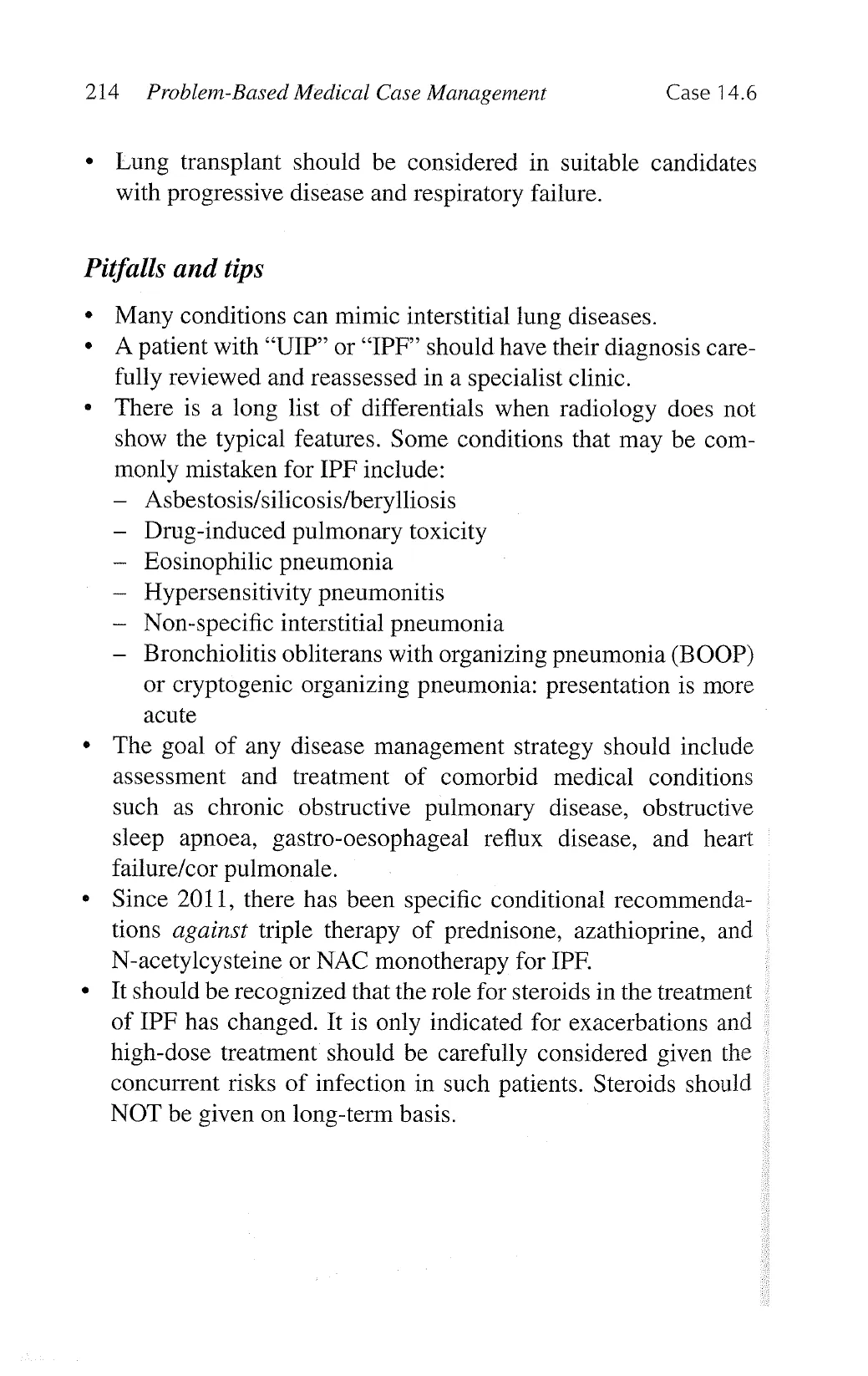
His chest X-ray is shown below:
View original page (figures / layout)
Page 22
216 Problem-Based Medical Case Management Case 14.7
Describe the radiological abnormalities.
Answer:
A right hilar mass consistent with diagnosis of lung cancer.
Arterial blood gas examinations while patient was breathing room
air showed pH 7.36, pO, 7.9 kPa and pCO, 5 kPa.
How would you describe this blood gas result?
Answer: Type I respiratory failure.
What is the next investigation of choice?
Answer: CT thorax with contrast (CT pulmonary angiogram)
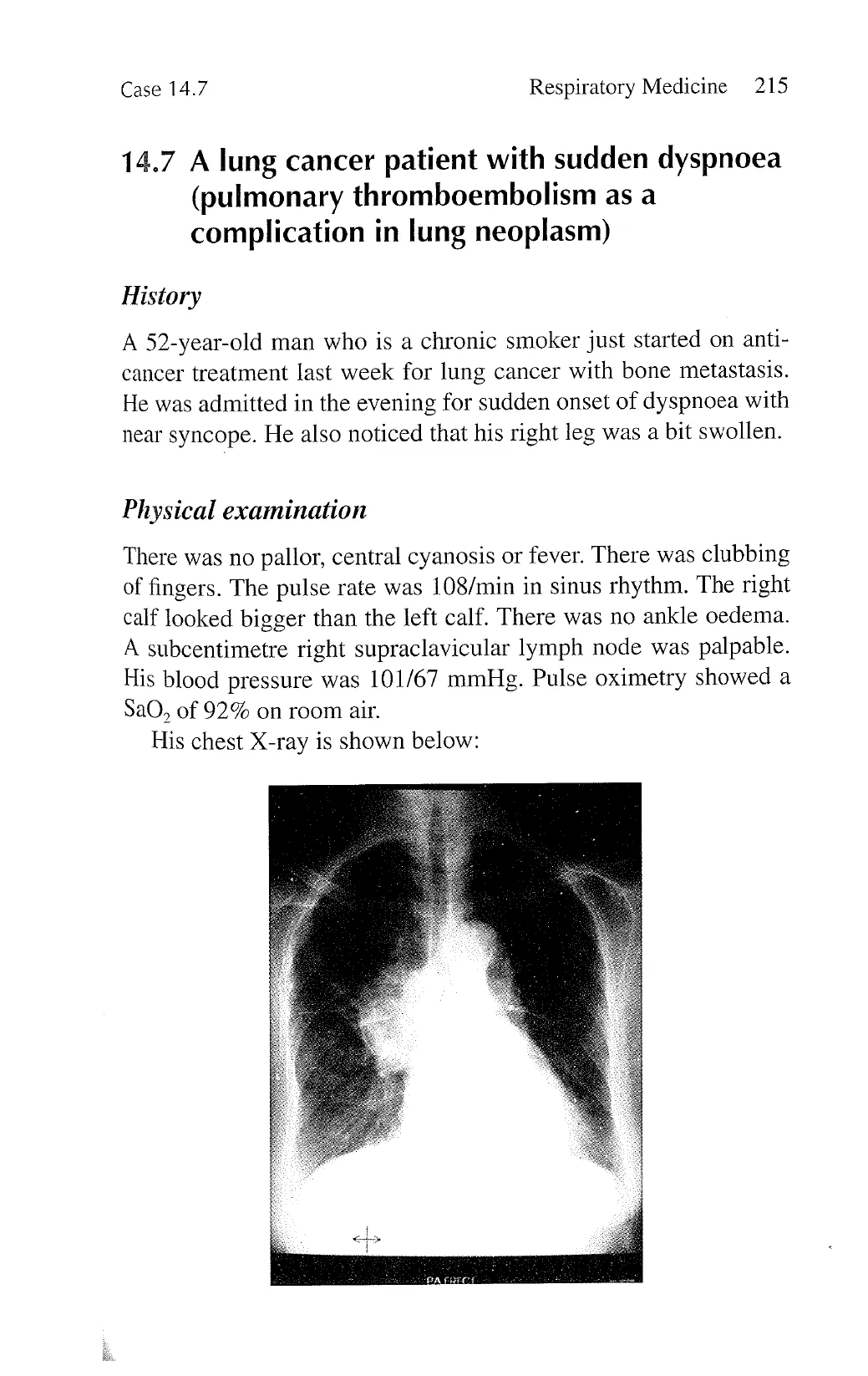
What is the radiological abnormality?
Answer:
Pulmonary thromboembolism shown as a filling defect in the pul-
monary vessel (red arrow).
What is the overall working diagnosis?
Answer:
Advanced stage lung cancer complicated with development of
deep vein thrombosis and pulmonary thromboembolism.
How should this patient be managed?
Answer:
1. Oxygen supplementation to relieve hypoxia.
2. Check blood including complete blood counts, liver and renal
function tests, and clotting profiles.
View original page (figures / layout)
Page 23
Case 14.7 Respiratory Medicine 217
3. Prescribe anti-coagulation, e.g., low-molecular-weight heparin.
Frequently asked questions:
1. How long should anti-coagulation therapy be maintained?
« Inpatients with active and persistent neoplasm complicated with
the development of venous thromboembolism, anti-coagulation
therapy should be maintained. Low-molecular-weight heparin
(LMWH) is the preferred form of maintenance anti-coagulation
therapy for patients with active neoplastic disease.
2. If bleeding occurs and patient cannot be given anti-coagula-
tion, is there another management option?
+ In patient with deep vein thrombosis of the lower limb, an
inferior vena caval filter could be installed to prevent further
thromboembolic phenomenon which could be life-threatening.
3. Could there be interactions between anti-coagulation and
anti-cancer therapy?
¢ There is no significant interaction between heparin with most
forms of chemotherapy used for the treatment of advanced
stage lung cancer.
There could be significant interaction between oral anti-
coagulant, i.e., warfarin, with EGFR-tyrosine kinase inhibitor
such that the anti-coagulant effects of warfarin will become
potentiated, which calls for careful titration of warfarin dosage,
usually in the form of dose reduction of warfarin when starting
on EGFR-TKIL.
4. How to handle the anti-coagulation if patient had to undergo
invasive investigation like bronchoscopy with anticipated need
for biopsy?
* In general, if a patient is on LMWH, stop it for at least 12 hours
before the procedure.
If a patient is on warfarin, stop it for at least 5 days before
the procedure, monitor INR and aim at INR < 1.5 before the
procedure, and switch over to low-molecular-weight heparin
until 12 hours before the procedure.
The clotting profile is checked again before the procedure.
View original page (figures / layout)
Page 24
218 Problem-Based Medical Case Management Case 14.7
5. Can we use the newer anti-coagulants such as tinzaparin,
or direct thrombin inhibitor (e.g., dabigatran or rivaroxaban)
for treatment of deep vein thrombosis and venous
thromboembolism?
« Instead of the conventional LMWH called enoxaparin, patient
can also be treated with a new type of LMWH called tinzaparin.
At the time of writing this book, the newer LMWH has been
shown to have comparable safety and efficacy profile as the
more conventional enoxaparin, but it has limited data on DVT/
VTE prophylaxis compared to enoxaparin.
After administration of LMWH, patients may be switched
over to oral direct anti-thrombin inhibitors (dabigatran or rivar-
oxaban) for maintenance (adjust the dosage according to renal
function). The handling of these newer oral direct anti-thrombin
inhibitors before bronchoscopy or surgery is also different from
warfarin, and these inhibitors could be stopped for 2 days before
the procedures (in subjects with impaired renal function, they
should be stopped for 4 days).
View original page (figures / layout)