Page 1
12
Neurology—Long Cases
Raymond Tak-Fai Cheung, Shu-Leong Ho, Windsor Mak,
and Koon-Ho Chan
12.1 Confused speech (embolic stroke due to
rheumatic mitral valve disease and atrial
fibrillation)
History
A 35-year-old right-handed woman presented with sudden onset
of confused speech and poor communication for 3 days. She was a
new immigrant from mainland China to Hong Kong and lived in an
apartment in a private housing estate, with her 60-year-old husband
and two teenage children from his first marriage. She was brought
up in a farming village, had not had any major medical illness in
the past, and was a non-smoker and non-drinker. However, her
exercise tolerance was not as good as her peers and so she could
not perform heavy manual farm work as much as others in her
family did. Three days ago, she was found to have confused speech
with difficulty in verbal communication. Her husband thought that
she had a flu and kept her at home. As there was no improvement,
she was brought to the accident and emergency department and
then admitted to a medical ward. Before the current episode, she
had complained of palpitations especially with exertion, and her '
exercise tolerance was limited to 2 flights of stairs. She was not
on oral contraceptive pills. She had no fever, flu-like symptoms,
constitutional upset, or motor weakness.
View original page (figures / layout)
Page 2
Case 12.1 Neurology 165
Physical examination
She was alert, conscious, and afebrile. Her blood pressure was
110/60 mmHg. She had a totally irregular, small volume pulse
with a rate of 90 per minute. All peripheral pulses were felt. The
apex was not displaced. There was a parasternal heave, but no
thrill was detected. On auscultation, the apical rate was 110 per
minute, the first heart sound was normal, the pulmonic component
of her second heart sound was loud, and two murmurs were heard
best at the apex. There was a grade 3/5 pansystolic murmur radi-
ating to the axilla, and another grade 3/5 mid-diastolic murmur
was louder with her lying in the left lateral position. Neurological
examination was difficult, as she failed to understand instructions.
Her spontaneous speech and naming of objects appeared normal.
Examination of her cranial nerves yielded nothing of note, and
muscle bulk, tone, and power in her limbs were entirely normal.
Her reflexes were symmetrical; both plantar responses were down-
going, coordination was normal, and sensations were intact. All
other systems were normal.
Questions
1. What are the diagnoses and on what grounds are they made?
* Cardiac: mixed mitral valve disease, pulmonary hypertension,
and atrial fibrillation.
Neurological: Wernicke’s aphasia (posterior part of the left
superior temporal gyrus) from a cardioembolic stroke. The
history of reduced exercise tolerance, palpitations (atrial fibril-
lation), mixed mitral valve disease, and sudden onset of fluent
aphasia are all consistent with this diagnosis.
2. What are the supportive physical findings for these
diagnoses?
* Signs of a cardiac problem include a pansystolic murmur at the
apex from mitral regurgitation, a mid-diastolic murmur with
postural accentuation from mitral stenosis, atrial fibrillation
from irregular pulse and a higher apical rate than peripheral
pulse rate, pulmonary hypertension from a parasternal heave,
and a loud pulmonic second heart sound.
View original page (figures / layout)
Page 3
166 Problem-Based Medical Case Management Case 12.1
.
3.
Signs of a stroke affecting Wernicke’s area include fluent
aphasia and otherwise normal neurological examination.
Clinically how do you determine the severity of mitral ste-
nosis versus mitral regurgitation in patients with mixed mitral
valve disease?
5.
Features of predominant mitral stenosis include: a small volume
pulse, an undisplaced tapping cardiac apex, a long diastolic
murmur, and signs of pulmonary hypertension.
Features of predominant mitral regurgitation include a dis-
placed cardiac apex and a palpable systolic thrill.
. How would you investigate the patient?
Blood tests should include complete blood counts, liver and
renal. function tests, coagulation tests, fasting glucose and
lipids. Neurologic investigations should include a plain com-
puted tomography (CT) brain to show any infarct over the
Wernicke’s area and to exclude haemorrhage, tumour, or other
lesions. A normal CT is possible, in which case magnetic reso-
nance imaging (MRI) stroke package may be more revealing.
Cardiologic investigations include assessment of heart size
and lung fields on CXR, ECG to document atrial fibrillation,
and an echocardiogram to assess mitral stenosis and regurgi-
tation, left atrial size, the presence of intra-cardiac thrombus
or vegetations on any valves. An MRI stroke package includes
brain MRI, brain magnetic resonance angiography (MRA), and
neck MRA.
What are the potential risks of rheumatic mitral valve
disease?
Systemic and cerebral embolization, infective endocarditis,
systemic and cerebral haemorrhage from long-term anti-coagu-
lation, heart failure, and pulmonary hypertension.
. How would you treat the patient?
The patient will require long-term anti-coagulation and speech
therapy.
What advice would you give to the patient?
She will need advice on diet, drug compliance, and regular
monitoring of her long-term anti-coagulation.
View original page (figures / layout)
Page 4
Case 12.2 Neurology 167
8. Where is the Wernicke’s area?
« It is located at the posterior part of the left superior temporal
gyrus. Lesion involving this area will lead to Wernicke’s or
receptive aphasia.
9. Would there be any concern about performing an MRI
examination if she had received a valvular surgery?
« The metallic artificial heart valve may or may not be compatible
with MRI examination.
Pitfalls and tips
* Failure to detect receptive aphasia
* Failure to elicit the cardiac signs
* Failure to link them together
12.2 Epilepsy with poor seizure control
History
A 35-year-old narcotic abuser and street-sleeper with an unre-
markable past medical history was admitted via the accident and
emergency department to a medical ward. He was found unarous-
able in a park. The ambulance officer noticed a few twitches in
his legs while transporting him. He regained consciousness before
arriving at the hospital.
Physical examination
Physical examination was unremarkable.
Investigations
He refused blood taking, but urine toxicology showed presence
of methadone. Brain CT was normal. Electroencephalography
showed a transient run of bilateral frontal sharpish discharges that
lasted for 2 seconds.
View original page (figures / layout)
Page 5
168 Problem-Based Medical Case Management Case 12.2
Clinical course
He was discharged after 7 days of observation. During this period,
he remained asymptomatic other than having some non-specific
giddiness and headache. Epilepsy was diagnosed in view of the
history of loss of consciousness associated with leg twitching
and the electroencephalograhic findings. Phenytoin was started.
A monthly disability allowance was also approved, though he
continued to work as a part-time taxi driver. Subsequently, he was
repeatedly admitted to the medical wards via the accident and
emergency department for seizures. He claimed that his attacks
were precipitated by hunger or bad weather. CT brain, electro-
encephalography, and Holter monitoring were performed during
some of these admissions, but did not show any abnormalities.
The patient always refused blood taking. He had several witnessed
seizures during hospitalization, which occurred when he was upset
or after quarrelling with the nurses. Sodium valproate, phenobar-
bitone, and clonazepam were added on top of phenytoin, but his
condition did not improve. In view of poor seizure control, he was
referred to the epilepsy clinic for workup of possible surgically
amenable epilepsy syndromes. Investigations including magnetic
resonance imaging and single photon emission computed tomogra-
phy scan were negative. Video-electroencephalographic telemetry
failed to detect any events after 14 days of recording. He refused
further invasive recordings. His medications were changed to a
combination of valproate, lamotrigine, topiramate, and gabapen-
tin. He insisted on continuing phenobarbitone and clonazepam,
because he thought these drugs “were helping him”, claiming such
treatment had improved his condition by 50%.
Further information
One day, he was arrested for illegal trading of medications at a
drugstore. All along he had been selling his supply of valproate,
lamotrigine, gabapentin, and topiramate in exchange for cash or
methadone. He shared his phenobarbitone and clonazepam with
his friends. He and the drugstore pharmacist were found guilty.
View original page (figures / layout)
Page 6
Case 12.2 Neurology 169
After release from prison, he still attended the hospital frequently
for episodic loss of consciousness. A video-electroencephalo-
graphic telemetry was repeated, which documented 18 events over
3 days. Analysis of the recordings showed that they were actually
pseudoseizures. All medications as well as the disability allowance
were discontinued. After that, he changed his complaint to recur-
rent abdominal pain.
Questions
1. What are epileptic seizures and epilepsies?
* An epileptic seizure is a brief and usually unprovoked stereo-
typed disturbance of behaviour, emotion, motor function or
sensation, which on clinical evidence results from a cortical
neuronal discharge.
* Epilepsy is diagnosed when there are recurrent unprovoked
seizures.
2. What are the differential diagnoses of epileptic seizures?
* Syncope.
* Metabolic encephalopathy, e.g., hypoglycaemia, hepatic
encephalopathy.
* Hyperventilation syndrome.
¢ Drop attacks.
* Sleep disorders, e.g., sleep walking, narcolepsy-cataplexy
syndrome.
* Movement disorders, e.g., tics, hemifacial spasm, startle,
choreoathetosis.
* Psychiatric conditions, e.g., pseudoseizures, panic attack.
Transient global amnesia.
Transient ischaemic attacks.
3. How are blood tests relevant to suspected epileptic seizures?
To identify/exclude metabolic causes of epileptic seizures.
To identify the metabolic consequences of generalized seizures,
e.g., raised muscle enzymes, neutrophilia, hyperprolactinaemia.
To identify/exclude metabolic encephalopathy.
To check anticonvulsant levels.
View original page (figures / layout)
Page 7
170 Problem-Based Medical Case Management Case 12.2
5.
. What mistakes are illustrated in the clinical course?
Inappropriate starting of anti-convulsant after one suspected
epileptic seizure.
Known narcotic abuser with positive urine test for metha-
done, making drug overdose a possible explanation of loss of
consciousness.
Driving is forbidden especially for people working as drivers.
Drug level has not been checked to ensure adequate dosing and
compliance prior to add-on therapy.
Other than common side effects such as drowsiness, cere-
bellar syndrome, and drug interactions, what are the specific
side effects of phenytoin, sodium valproate, lamotrigine, and
topiramate?
.
Phenytoin: gum hypertrophy, acne, hirsutism, lupus-like syn-
drome, Stevens-Johnson syndrome, agranulocytosis.
Sodium valproate: alopecia, tremor, weight gain, dyspepsia,
hepatotoxicity, hyperammonaemia, acute pancreatitis.
Lamotrigine: skin rash.
Topiramate: paraesthesia, anorexia, weight loss.
Pitfalls and tips
Limb twitching is quite common in non-epileptic conditions
that cause impairment of consciousness.
An erroneous diagnostic label of epilepsy can have profound
consequences.
The main purpose of investigating a patient with seizure(s) is to
look for an underlying brain lesion or other secondary causes.
Epilepsy and seizure are diagnosed by history or clinical obser-
vation. The finding of EEG abnormalities is not specific for
epilepsy.
Non-compliance is a common cause of refractory epilepsy.
Pseudoseizure is a common cause of suspected refractory
epilepsy.
Manipulative behaviour as well as sick role can often be rein-
forced by financial gains.
View original page (figures / layout)
Page 8
Case 12.3 Neurology 171
+ Some anti-convulsants can be used as drugs of abuse.
« It is important to know the general and specific side effects of
common anti-convulsants.
12.3 Diplopia on reading (myasthenia gravis with
thyrotoxicosis)
History
A 23-year-old woman presented to a private practitioner with a
6-week history of generalized malaise, weight loss of 8 pounds,
fine hand tremor, and increased sweating. She was a cashier, and
her past health was good. She was single, did not take regular medi-
cations, and had no family history of neurological disease. She was
diagnosed to have thyrotoxicosis with blood free T4 level raised to
38 U/L and TSH level suppressed to 0.22 U/L. She responded to
treatment with carbimazole, which was successfully tailed off after
1 year. However, 9 months later, she noticed increased fatigability
of all four limbs and double vision after prolonged reading. She
had no dysarthria or dysphagia, but her exercise tolerance dete-
riorated from being essentially unlimited to climbing 3 flights of
stairs (limited by limb fatigability). Her symptoms were worse in
the evening.
Physical examination
Examination revealed a clinically euthyroid woman with coherent
speech and mild right partial ptosis that worsened on prolonged
Upward gaze. She complained of diplopia on bilateral horizontal
gaze with partial impairment of right eye abduction and left eye
adduction noted upon repeated eye movements; pupils were equal
and reactive to light. Other cranial nerves were intact. Muscle tone
Was normal with grade 4/5 proximal muscle weakness. Tendon
reflexes, gait, and sensation were normal, and she had no cerebel-
lar signs.
View original page (figures / layout)
Page 9
172 Problem-Based Medical Case Management Case 12.3
Investigations
Tensilon test (intravenous edrophonium injection) was positive,
with marked improvement of ptosis and resolution of diplopia.
Serum acetylcholine receptor antibody test was positive. Computed
tomography scan of the thorax did not reveal any thymic mass.
Repetitive nerve stimulation over her trapezius at 3 Hz revealed a
20% decremental response.
Questions
1. After 6 months of mestinon at 30 mg tds, she noticed
increased proximal muscle weakness and moderate dysphagia.
How do you manage her?
* Mestinon dose could be stepped up for an inadequate response.
Systemic corticosteroids and steroid sparing drugs, such as
azathioprine, were not started due to a concern of potential
long-term adverse effects, including risk of lymphoproliferative
diseases.
2. What are the non-drug alternatives for myasthenia gravis?
* Thymectomy should be advised, and significant improvement
in the absence of a thymoma can occur after thymectomy.
¢ Plasmapheresis has been used in intractable cases, but the
benefit is temporary.
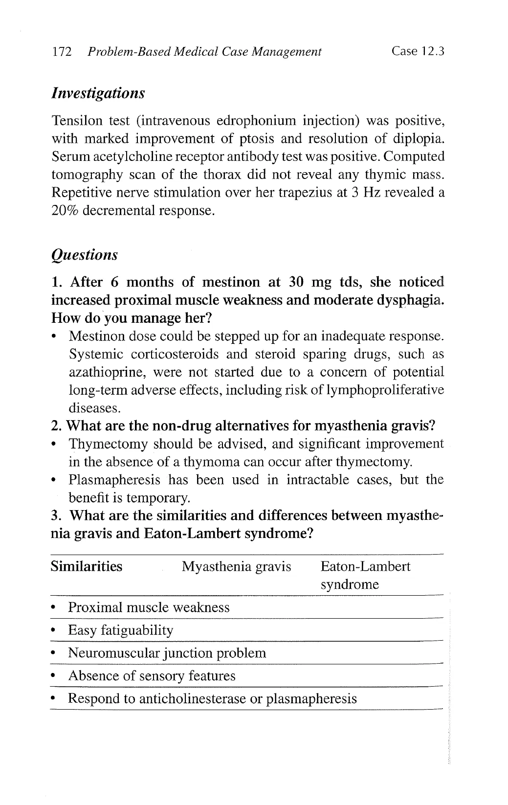
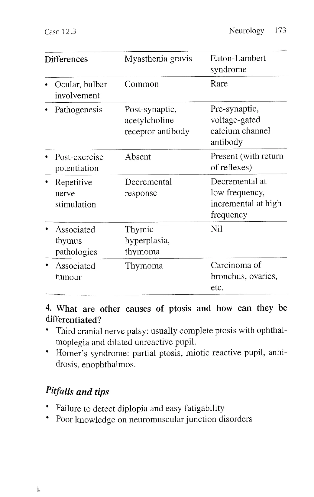
3. What are the similarities and differences between myasthe-
nia gravis and Eaton-Lambert syndrome?
Similarities Myasthenia gravis Eaton-Lambert
syndrome
¢ Proximal muscle weakness
» Easy fatiguability
¢ Neuromuscular junction problem
* Absence of sensory features
* Respond to anticholinesterase or plasmapheresis
View original page (figures / layout)
Page 10
Case 12.3 Neurology 173
Differences Myasthenia gravis Eaton-Lambert
syndrome
¢ Qcular, bulbar Common Rare
involvement
Pathogenesis
Post-synaptic,
acetylcholine
receptor antibody
Pre-synaptic,
voltage-gated
calcium channel
antibody
* Post-exercise Absent Present (with return
potentiation of reflexes)
* Repetitive Decremental Decremental at
nerve response low frequency,
stimulation incremental at high
frequency
* Associated Thymic Nil
thymus hyperplasia,
pathologies thymoma
* Associated Thymoma Carcinoma of
tumour
bronchus, ovaries,
ete.
4. What are other causes of ptosis and how can they be
differentiated?
* Third cranial nerve palsy: usually complete ptosis with ophthal-
moplegia and dilated unreactive pupil.
* Horner’s syndrome: partial ptosis, miotic reactive pupil, anhi-
drosis, enophthalmos.
Pitfalls and tips
* Failure to detect diplopia and easy fatigability
* Poor knowledge on neuromuscular junction disorders
View original page (figures / layout)
Page 11
174 Problem-Based Medical Case Management Case 12.4
12.4 Tuberculous meningitis complicated
by stroke
History
A 30-year-old lady with swinging fever, night sweats, chills, and
rigors for 4 weeks presented acutely with disorientation. She had
received in vitro fertilization for primary infertility due to fallopian
tube blockage, using human chorionic gonadotrophin. She had
premature rupture of her amniotic membrane at 18 weeks of gesta-
tion, and was given 2 intravenous injections of betamethasone for
fetal lung maturation. Emergency caesarean section was carried
out for fetal bradycardia at 29 weeks of gestation. Several days
after delivery, her symptoms started. There was no joint pain, skin
rash, leg swelling, dysuria, cough, chest or abdominal discomfort,
or vaginal bleeding. Physical examination was normal. Empirical
broad-spectrum intravenous antibiotics were given. She remained
unwell, and her high swinging fever persisted. One month post-
partum, she had acute onset of confusion and disorientation, and
developed partial drooping of her right eyelid. Her antibiotic
regimen was adjusted to cover possible listeriosis. Her condition
deteriorated further, and she developed sudden-onset left hemi-
paresis. Anti-tuberculous treatment with steroids (dexamethsone)
was added empirically to cover tuberculous meningoencephalitis.
Aspirin was also added for her stroke. She gradually improved
over several months but with residual disability.
Physical examination
She was afebrile. There was no palpable lymph node or skin
rash. She was fully conscious and orientated. There was no neck
stiffness. She had left homonymous hemianopia, right partial
ptosis, and a small reactive right pupil. Fundoscopic examination
was normal. She required a wheel-chair for ambulation. Her left
upper limb was flexed at her elbow. Her left arm and leg were
weak (power of grade 3/5 to 4/5), with spasticity and hyperreflexia.
Her plantar response was upgoing on the left and down-going on
View original page (figures / layout)
Page 12
Case 12.4 Neurology 175
the right. Her peripheral sensations were intact, but she had left-
sided sensory neglect. She was in sinus rhythm, and examination
of her cardiovascular system did not reveal any carotid bruit or
heart murmur. Her blood pressure was 130/80 mmHg. Chest and
abdominal examinations were normal.
Investigations
CXR and ultrasound examination of her abdomen and pelvis were
normal. Repeated smear and culture of her blood, urine, and wound
swab were negative. Her echocardiogram was normal. Shortly
after the development of confusion and disorientation, an urgent
CT brain had revealed a large hypodense lesion in her right pari-
eto-temporal region compatible with a cerebral infarction. CSF
examination showed an opening pressure of 34 cm (water), CSF
protein 3.79 g/L, CSF glucose 2.1 (serum glucose 6.4), CSF white
cell count 30 (lymphocytes 56%, neutrophils 40%, monocyte 4%),
RBC 11, and Gram stain negative. One week after lumbar punc-
ture, polymerase chain reaction was positive for Mycobacterium
tuberculosis DNA in her CSE.
Questions
1. What is the most likely cause for her fallopian tube blockage?
* Tuberculous salpingitis.
2. Why did she develop persistent fever after delivery of her
baby?
* Reactivation of tuberculosis with development of tuberculous
meningitis.
3. From the CSF examination results, apart from TB menin-
gitis, what other diseases could give a similar clinical picture?
* Partially treated bacterial meningitis, fungal meningitis.
4. What empirical antibiotic regimen was most likely given
initially, and what antibiotic change would occur to cover for
listeriosis?
* Intravenous high-dose penicillin G and a third-generation
cephalosporin; high-dose ampicillin instead of penicillin G.
View original page (figures / layout)
Page 13
176 Problem-Based Medical Case Management Case 12.4
5. What is the possible mechanism for developing her cerebral
infarction?
¢ Thrombosis secondary to arteritis.
6. What are the other complications of tuberculous meningitis
and how are they treated?
¢ Hydrocephalus (shunting), cranial nerve palsies (steroids),
tuberculoma (steroids, anti-inflammatory drugs), spinal block
(steroids), tuberculous brain abscess (surgical drainage); anti-
tuberculous chemotherapy is required in all cases.
7. What anti-tuberculous regimen was most likely started, and
how long should she remain on the treatment?
¢ Isoniazid with vitamin B6 (pyridoxine), rifampicin, ethambu-
tol, pyrazinamide; for 9 to 12 months.
8. What are the side effects of each of the drugs used in the
regimen, and, if appropriate, what precautions would you
take to prevent these side effects or minimize their impact on
the patient?
¢ Isoniazid: hepatitis (desensitization), peripheral neuropathy
(vitamin B6); rifampicin: drug interactions (review drug chart),
hepatitis (desensitization), fever and vasculitis (cessation),
nausea and vomiting (daily); ethambutol: optic neuritis (regular
monitoring by ophthalmologist), hepatitis (desensitization);
pyrazinamide: hepatitis (desensitization), gout (colchicine and
allopurinol).
9. Why was dexamethasone given with the anti-tuberculous
treatment, and under what other circumstances would you add
steroids to the treatment?
e For anti-inflammatory effects in Medical Research Council
Stage II or III disease: lethargy, prominent meningeal irritation,
cranial nerve palsies, convulsion, paralysis, stupor, coma.
10. Which of these anti-tuberculous drugs can be safely used in
pregnancy with a minimal amount of teratogenicity?
* Isoniazid with vitamin B6 (pyridoxine), rifampicin, ethambutol.
View original page (figures / layout)
Page 14
g
Case 12.4 Neurology 177
Pitfalls and tips
+ Difficult to differentiate coexisting left homonymous visual
field loss (lesion affecting her right optic radiation) and left
sensory neglect (lesion affecting her non-dominant right pari-
etal lobe)
« Failure to associate tuberculous meningitis with stroke
* Poor knowledge in management of tuberculous infections
View original page (figures / layout)