Page 1
11 |
Neurology—Short Cases
Shu-Leong Ho, Raymond Tak-Fai Cheung, Windsor Mak,
Kay-Cheong Teo, and Koon-Ho Chan
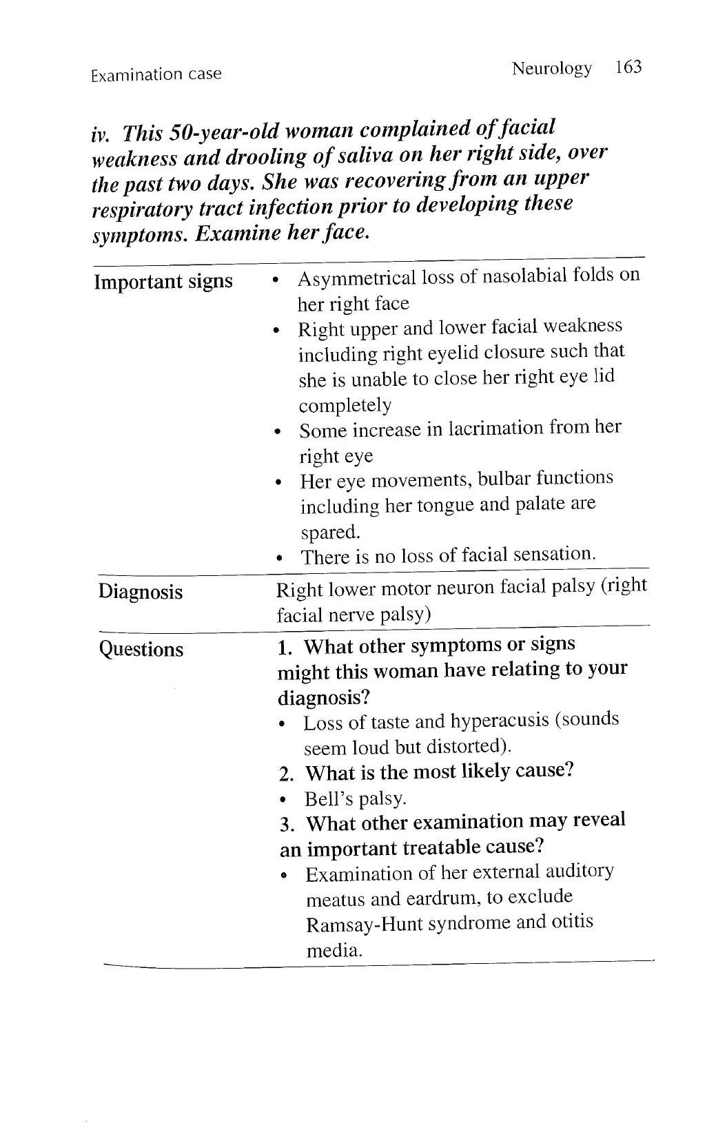
Overview
Elucidate the location and nature of the lesion in the nervous
system.
Assessing higher mental functions and discovery of any cranial
nerve deficits, pyramidal, cerebellar, extrapyramidal signs or
sensory disturbances can all help to locate the lesion.
Age of the patient, timing and mode of onset, pattern of progres-
sion, and response to empirical treatment may help to indicate
the nature of the lesion.
Examination of higher mental function
Assess mood, attention, orientation, language, memory (short-
term and long-term memory), calculation, abstract thought,
spatial, and visual perception.
Check orientation in time, place, and person.
Assess for comprehension, expression, and repetition.
For comprehension of language, ask patient to follow simple
written or verbal commands, e.g., close your eyes, lift up your
left arm.
For expression of language, ask patient to name objects.
Suggestions may be given if there is difficulty.
For repetition, ask patient to repeat a phrase.
View original page (figures / layout)
Page 2
Neurology 143
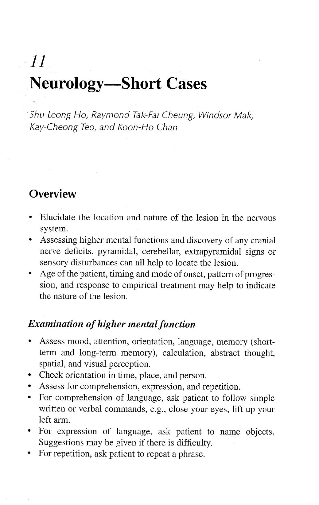
Ask patient to repeat three objects such as apple, table, and
penny, and then after 5 minutes ask for recall of the same
objects (short-term memory).
Ask patient to recall their telephone number, home address,
distant events or names of famous persons (long-term memory).
To test for calculation, ask patient to subtract 7 from 100, and 7
from the results thereafter.
Ask patient to copy 2 intersecting pentagons; write the numbers
on a round clock-face and draw the hands for a given time.
Ask patient to name the number drawn on the palm of either
hand or recognize coins of various denominations placed in
either hand.
To test for sensory inattention, touch the patient on each upper
limb individually and simultaneously, with the patient’s eyes
open and closed. Ask patient to name the side of the upper limb
being touched. After ensuring that there is no visual field loss,
test for visual inattention by asking patient to point to your
finger which moves on either side of his visual field.
Give a scenario to the patient and assess if there are any irra-
tional or concrete thoughts. Ask patient the meaning of various
proverbs. Ask patient for similarities between different objects
with the same utility (e.g., boat and car, banana and apple).
Examination of cranial nerves
.
Ask for loss of smell or taste.
Inspect for scars, facial asymmetry, deformities, pupil size,
proptosis.
Check visual acuity of each eye using Snellen chart. If the
patient is unable to see the chart, use finger counting, hand
movement, and light from a torch.
Compare the visual field of each eye on direct confrontation.
Check direct and consensual pupillary responses, and test for
the accommodation reflex by looking at a distant object fol-
lowed by a near object.
Look at both fundi.
View original page (figures / layout)
Page 3
144 Problem-Based Medical Case Management
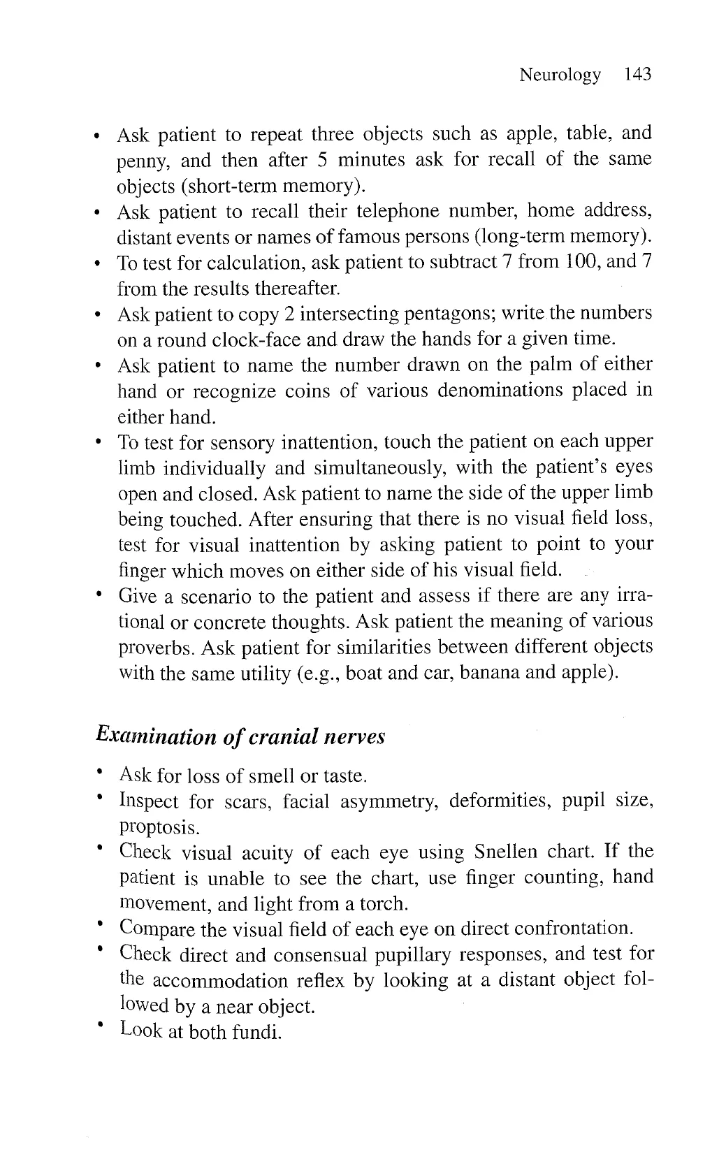
Examine eye movements for dysconjugate gaze and nystagmus
(horizontal or vertical).
Ask patient to bite and open the mouth against resistance.
Perform the jaw reflex.
Check for facial sensation.
Use a clean cotton wool to examine for corneal reflexes.
Ask patient to close both eyes tightly and look upwards (look
for asymmetry of the forehead skin creases), show teeth and
blow up the cheeks.
Perform the Weber’s and Rinne’s test on both ears using a
tuning fork (256 Hz).
Listen for dysarthria, and test articulation by asking the patient
to repeat standard words (e.g., British constitution, baby hip-
popotamus) or sentences.
Look for uvula movement and deviation, when the patient is
asked to say “ahhh”.
Perform the gag reflex.
Inspect the relaxed tongue for muscle bulk and fasciculation.
Ask patient to protrude the tongue, move the tongue from side
to side or in/out, and protrude it into either cheek.
Examination of limbs
Observe: gait (during usual walking and if appropriate heel-toe
walking), posture, muscle bulk, fasciculations, spontaneous or
involuntary movements.
Examine tone with the patient completely relaxed.
Test strength of specific groups of muscles across single joints.
Elicit limb reflexes with a tendon hammer, with the patient
completely relaxed. If the reflex is absent or diminished, ask
the patient to perform Jendrassik manoeuvres for reinforcement
(e.g., clenching the teeth, interlocking of fingers in a hook-like
form). If the reflex is brisk, test for clonus. Using the tendon
hammer requires a lot of practice. The best effect comes from a
wrist action to swing the hammer.
View original page (figures / layout)
Page 4
Case 11.1 Neurology 145
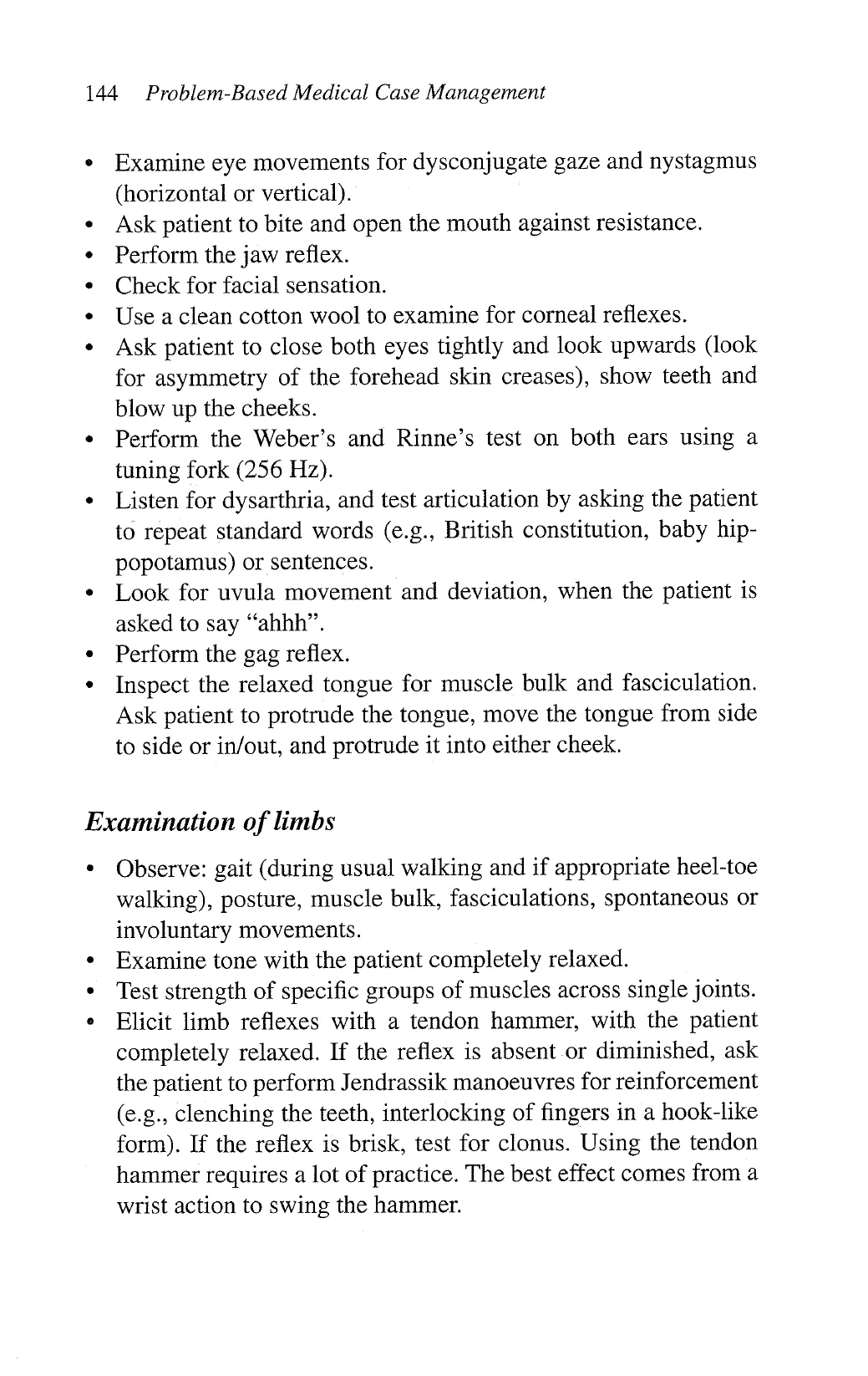
Elicit for plantar response.
Assess for sensory deficits from two perspectives:
— Modality (i.e., dorsal column or spinothalamic loss).
— Distribution (dermatomal or glove/stocking loss).
Test for bradykinesia and cerebellar signs if indicated.
11.1 Cerebellar syndrome
Classical signs
Truncal ataxia or limb ataxia with wide-based ataxic gait;
unable to perform heel-toe walking despite normal strength
Nystagmus on horizontal or vertical conjugate gaze
Dysarthria, typically slow and slurred speech
Intention tremor
Past-pointing
Dysdiadochokinesia
Additional signs may be evident depending on the cause of the
cerebellar syndrome, e.g., upper motor neuron signs in multiple
sclerosis, wasting/lymphadenopathy/clubbing in paraneoplastic
syndrome.
Causes
Cerebellar degeneration: idiopathic, paraneoplastic, familial
Toxins and drugs: alcohol, anticonvulsants
Cerebellar infarct, haemorrhage, tumour (primary or metastases)
Important investigations
Computed tomography/magnetic resonance imaging of brain
Serum for paraneoplastic anti-neuronal autoantibodies
* Cerebrospinal fluid analysis to exclude infection and
inflammation
View original page (figures / layout)
Page 5
146 Problem-Based Medical Case Management Case 11.2
Discussion
Neurological features may precede detection of malignancy.
Common associated malignancies are carcinoma of lung,
breast, and female genital tract.
Pitfalls and tips
Exclude muscle weakness before assessing for ataxia. In the
presence of significant limb weakness, intention tremor and
past pointing are difficult to interpret.
Sensory ataxia typically worsens with eyes closed (positive
Romberg’s test):
11.2 Chronic inflammatory demyelinating
polyneuropathy (CIDP)
Classical signs
Progression over 8 weeks
Symmetrical flaccid tone, proximal and distal muscle wasting,
and weakness in all the limbs
May develop foot-drop in more severe cases
Diminished or absent limb reflexes
May have absent plantar response
Glove and stocking pattern of sensory loss
Additional signs of iatrogenic complications: Cushingoid
features because of long-term corticosteroid therapy, surgical
scar posterior to lateral malleolus after sural nerve biopsy for
diagnosis
Cause
.
Immune-mediated demyelination of the peripheral nerves
[
View original page (figures / layout)
Page 6
P
Case 11.2 Neurology 147
Important differential diagnoses
Diabetic distal symmetric sensorimotor polyneuropathy
Acute or subacute forms of Guillain-Barre syndrome
Hereditary motor sensory neuropathies (Charcot-Marie-Tooth
disease), hereditary neuropathy with liability to pressure palsies
(HNPP)
Paraproteinemic or paraneoplastic neuropathies
Neuropathies associated with connective tissue diseases
Drugs or toxins (mercury, arsenic, N-hexanes, chloroquine,
amiodarone, vincristine)
Vitamin B12 deficiency
HIV- or CM V-associated inflammatory neuropathy
Important investigations
Electrophysiological studies
Cerebrospinal fluid analysis (elevated protein level but no
pleocytosis)
Screen for paraproteins, autoimmune connective tissue dis-
eases, malignancies.
Sural nerve biopsy (reduction in myelinated fibres, endoneurial
mononuclear cell infiltration, onion bulb formation and other
remyelinating features)
Discussion
Differential diagnosis and investigations of peripheral neuropa-
thies. CIDP has a protracted course (over 8 weeks), progressive
or relapsing-remitting in nature.
Treatment: corticosteroids, immunosuppressants, intravenous
immunoglobulins, plasmapharesis
Pitfalls and tips
Look for generalized and symmetrical ngture of the signs in
the limbs.
View original page (figures / layout)
Page 7
148 Problem-Based Medical Case Management Case 11.3
Pes cavus indicates an earlier. pathogenic onset, developing in
childhood (e.g., inherited causes) and is not present in late-
onset neuropathies.
Respiratory and bulbar muscles may be involved in advanced
cases.
11.3 Lateral medullary syndrome
Classical signs
Ipsilateral signs:
— Dysphagia, hoarseness of voice due to vocal cord paralysis,
loss of gag reflex, soft palate paralysis, with uvula deviation
when patient says “ahhh” (IX, X nucleus)
— Horner’s syndrome (descending sympathetic fibres)
— Loss of all sensory modalities of face (descending sensory
tract and nucleus of V nerve)
— Cerebellar ataxia with nystagmus towards the side of lesion
(inferior cerebellar peduncle and cerebellar connections)
Contralateral loss of pain and temperature sensation below
the face
Additional signs depending on cause: risk factors of stroke
(e.g., xanthelasma, corneal arcus, nicotine stains, hypertension,
diabetic features), recent neck trauma
Causes
Usually brainstem infarction from local thrombotic occlusion,
arterial thromboembolism (e.g., vertebral artery dissection) or
cardioembolism
Brainstem haemorrhage
Acute brainstem demyelination: multiple sclerosis
Leukoencephalopathy with brainstem involvement (hyperten-
sion, drugs, autoimmune diseases)
View original page (figures / layout)
Page 8
Case 11.3 Neurology 149
Important differential diagnoses
+ Acute vestibular neuronitis or benign positional vertigo,
in which focal neurological signs (except for nystagmus) are
absent
Important investigations
«+ Computed tomography/magnetic resonance imaging of brain
+ Exclude vascular risk factors
Discussion
+ Most cases are due to occlusion of the vertebral artery and its
lateral medullary penetrating arteries. Posterior inferior cer-
ebellar artery occlusion accounts for < 15% of cases. Hence,
cerebellar infarction is uncommon in this syndrome. The cer-
ebellar signs are due to lesion affecting the inferior cerebellar
peduncle and its connections in the brainstem.
* Anti-platelet therapy remains the primary treatment for
ischaemic stroke. The use of anti-coagulation is controversial
in arterial dissection but 3 to 6 months of anti-coagulation is
sometimes prescribed, or if there is progression of thrombosis
despite anti-platelet therapy.
Reperfusion therapy in acute ischaemic stroke
Pitfalls and tips
* If patient has occipital headache and neck pain, consider verte-
bral artery dissection.
If contralateral weakness is present (indicating involvement
of the corticospinal tract located more medially), it indicates
a more extensive lesion, such as haemorrhage, demyelination,
Or tumour,
View original page (figures / layout)
Page 9
150 Problem-Based Medical Case Management Case 11.4
11.4 Amyotrophic lateral sclerosis (ALS)
Classical signs
Thin, may be breathless from respiratory muscle weakness
Muscle wasting (may be asymmetrical) with fasciculation and
weakness in the limbs and tongue. Typically a mix of upper
and lower motor neuron lesion signs. Depending on the pre-
dominant lesion, reflexes may be brisk, normal, absent, and
asymmetrical. Plantar responses may be upgoing, down-going
or absent.
Palatal paralysis and dysarthria with nasal speech (bulbar or
pseudobulbar palsy).
Eye movements are preserved with a normal range of horizontal
and vertical gaze.
Absence of sensory signs
Important differential diagnoses
Cervical myelopathy with multiple radiculopathy: presence of
sensory deficits and other signs, e.g., Hoffman’s sign, inverted
supinator jerks; tongue and bulbar muscles not affected
Spinal muscular atrophy: differentiated from ALS by the
absence of upper motor neuron signs, more benign clinical
course, and electromyogram findings
Motor predominant neuropathy: lower motor neuron lesion
signs, with or without sensory features
Poliomyelitis: fasciculation in the acute stage; wasting, possible
contractures, and other lower motor neuron lesion signs in the
chronic stage
Muscular dystrophy: presenting with distal or asymmetrical
wasting in specific muscle groups, typically the shoulder or calf
muscles; no sensory deficit
Polymyositis: muscle fasciculations and weakness without any
sensory features, especially affecting the proximal muscles;
early wasting unusual
View original page (figures / layout)
Page 10
Case 11.4 Neurology 151
- Benign fasciculation syndrome: persistent muscle fascicula-
tions with no identified cause; diagnosis by exclusion.
« Thyrotoxicosis: muscle wasting and fasciculation possible
Important investigations
+ Electromyogram: chronic denervation and reinnervation with
high amplitude motor unit action potentials. Spontaneous
muscle activity consisting of positive sharp waves, fibrillations
and complex repetitive discharges.
*+ Sensory and motor nerve conduction parameters are normal in
early stages, but compound motor action potential amplitude
may be reduced in severely wasted muscles.
+ Serum creatine kinase level is usually normal or slightly raised.
*+ Magnetic resonance imaging of the cervical spine to exclude
cervical cord pathology
Discussion
* Subtypes: primary lateral sclerosis (mainly upper motor neuron
deficits); progressive muscular atrophy (mainly lower motor
neuron deficits); progressive bulbar palsy (bulbar with upper
and lower motor neuron deficits)
Prognosis: 50% die usually from respiratory failure or aspira-
tion pneumonia within 3 years; 90% die within 5 years.
Riluzole has a survival benefit of about 3 months.
Palliative care, nutrition, e.g., naso-gastric or percutaneous gas-
trostomy feeding
Consider intermittent ventilatory assistance or long-term
mechanical ventilation. Depression is frequent. Consider coun-
selling, family and social support, and antidepressants.
Pitfalls and tips
Look for fasciculations with the muscles at rest. Tap the muscles
gently if none are observed; avoid rushing to tap muscles
without properly observing them first.
View original page (figures / layout)
Page 11
152 Problem-Based Medical Case Management Case 11.5
¢ Ask the patient to open the mouth without protruding the tongue
and inspect for fasciculation. A normal tongue, when protruded
may appear “fasciculating”.
11.5 Multiple sclerosis
Classical signs
¢ Optic neuritis: impaired visual acuity, afferent pupillary defect,
with diminished direct and consensual pupillary response
to light
¢ Internuclear ophthalmoplegia: dysconjugate gaze palsy which
results in impaired adduction in the ipsilateral eye with nystag-
mus in the contralateral abducting eye on horizontal gaze
¢ Spastic paraparesis: unsteady stiff gait, characterized by
increased tone and associated weakness in both lower limbs.
Typically, brisk knee and ankle reflexes are encountered and the
Babinski’s sign is present bilaterally. Reduced sensation in all
modalities in both lower limbs, particularly in association with
a sensory level.
» Cerebellar signs: dysarthria, intention tremor and past-pointing
on finger-nose test. Impaired rapid repetitive motion of the
hands and feet (dysdiadochokinesia).
Important differential diagnosis
» Neuromyelitis optica (NMO)
e Central nervous system manifestation of rheumatological
diseases
Important investigations
* Blood for NMO-IgG
* Magnetic resonance imaging of brain, brainstem, and spinal cord
show multiple demyelinating plaques, typically in the periven-
tricular regions, corpus callosum, and centrum semiovale
View original page (figures / layout)
Page 12
Case 11.6 Neurology 153
« Lumbar puncture for cerebrospinal fluid analysis may show
presence of oligoclonal bands.
+ Visual evoked response may be delayed.
Discussion
« Multiple sclerosis is a demyelinating disease of the central
nervous system, which demonstrates lesion disseminated in
time and location. Typically, it presents with relapsing-remit-
ting symptoms, but other clinical patterns occur, e.g., primary
(progression of disease from onset) or secondary progressive
(initial relapsing-remitting followed by progression), or pro-
aressive relapsing (progressive from onset and during the inter-
vals between each relapse).
* Treatment: pulsed IV corticosteroids for acute relapse, immu-
nomodulatory drugs to modify the course of the disease
(i. interferon beta; ii. oral agents: fingolimod, dimethyl fuma-
rate, teriflunomide; iii. monoclonal antibodies: natalizumab)
Pitfalls and tips
* Examine the fundus in a darkened room. Look for the red reflex.
Zoom in slowly with the fundoscope, and orientate by locating
the optic disc first.
Think of internuclear opthalmoplegia when nystagmus is found
with dysconjugate gaze.
Demyelination of the peripheral nervous system, by definition,
is not multiple sclerosis.
11.6 Parkinson’s disease
Classical signs
* Stooped posture (flexed at the trunk), thin, masked (expres-
sionless) facies, soft monotone voice, drooling of saliva, greasy
facial skin, typically in elderly patient
View original page (figures / layout)
Page 13
154 Problem-Based Medical Case Management Case 11.6
Stiff, shuffling or festinant (increasing rapid small steps with
centre of gravity shifted forward) gait, lacking arm swing
Resting tremor, typically asymmetrical, “pill-rolling”, affecting
the hands
Cog-wheel rigidity more prominent in the limbs compared to
the trunk
Bradykinesia (slow in initiating movement), may “freeze”
during walking or turning
Positive glabellar tap sign (unreliable)
Power, sensation, and deep tendon reflexes are preserved.
Important differential diagnoses
Multisystem atrophy (MSA): 2 clinical subtypes, i.e., parkin-
sonism with i) predominant pyramidal features (MSA-P), or
ii) with predominant cerebellar ataxia (MSA-C). Autonomic
failure is an important feature and occurs early. Bilateral vocal
cord palsy causing stridor may be life-threatening.
Progressive supranuclear palsy: vertical gaze palsy (unable to
look up or down), prominent dysarthria, truncal rigidity. These
patients lack resting tremor.
Lewy body dementia: parkinsonism with hallucinations and
psychosis occur in early stages of the disease, and eventual
dementia.
Wilson’s disease: Kayser-Fleischer ring best seen under slit-
lamp examination, younger age at onset; consanguineous
parents, prominent dystonia and patient may develop limb con-
tractures, psychosis and jaundice.
Cerebral small vessel disease and normopressure hydrocepha-
lus may cause parkinsonism affecting the lower limbs more
than upper limbs.
Benign essential tremor: bilateral postural tremor, worse when
performing a task. These patients may have head titubation or
dystonia, but without parkinsonian signs.
View original page (figures / layout)
Page 14
Case 11.7 Neurology 155
Iinportant investigations
+ Family and drug history (e.g., long-term neuroleptics, antihista-
mines, and illicit drugs)
« Copper studies, slit-lamp examination in patient with young
age of onset
+ Magnetic resonance imaging of the brain if atypical features
(e.g., early onset cerebellar, pyramidal or autonomic distur-
bance or dementia) are present.
Discussion
+ Pathology: degeneration of dopaminergic nigrostriatal neurons,
resulting in progressive loss of dopamine supply. Associated
with Lewy bodies.
+ Principles of drug treatment and their complications
Pitfalls and tips
* Examine for atypical features, e.g., gaze palsy, cerebellar and
pyramidal signs, early dementia or psychotic features, promi-
nent truncal rigidity, prominent autonomic failure to help differ-
entiate other forms of parkinsonism from Parkinson’s disease.
* A typical pill-rolling, resting tremor, and asymmetrical par-
kinsonian features in the upper limbs are helpful indicators of
Parkinson’s disease.
* Ask the patient to walk. Reduced arm swing is an early sign
for Parkinson’s disease and walking may “bring out” resting
tremor.
11.7 Polymyositis/Dermatomyositis
Classical signs
* Symmetrical proximal limb and neck weakness, muscle
tenderness
View original page (figures / layout)
Page 15
156 Problem-Based Medical Case Management Case 11.7
Muscle bulk is usually spared, especially in early stages.
Sensation is intact.
Dermatomyositis if the skin is involved; typically there is a
purple heliotrope in a butterfly distribution over the cheeks and
eyelids that blanches with pressure. Skin manifestations may
precede muscle weakness.
Additional signs: Cushingoid features from corticosteroid treat-
ment, cachectic appearance, lymphadenopathy, radiotherapy
marks in patients with underlying malignancy
Important differential diagnoses
Polymyalgia rheumatica, typically affects elderly women, and
may be associated with temporal arteritis. However, polymyal-
gia rheumatica and temporal arteritis are rare in Chinese.
Limb girdle muscular dystrophy (associated with early wasting
in shoulder or pelvic girdle muscles)
Myasthenia gravis: weakness is typically fatiguable and
involves the eyes.
Patients with systemic lupus erythematosus can have rash and
myositis, classically a photosensitive butterfly facial rash.
Important investigations
o
Increased serum creatine kinase level
Electromyography shows myopathic changes.
Muscle biopsy shows scattered fibre necrosis and perivascular
inflammatory infiltration.
Discussion
Associated underlying malignancy, e.g., cancers of ovaries,
lung, pancreas, breast, gastrointestinal tract, nasopharynx and
non-Hodgkin’s lymphoma. Malignancy may develop before
muscle weakness.
Often follows a relapsing-remitting course
View original page (figures / layout)
Page 16
Case 11.8 Neurology 157
« Treatment: corticosteroids, immunosuppressants (azathioprine
and cyclophosphamide), and intravenous immunoglobulins
may be useful.
Pitfalls and tips
* Proximal weakness manifests as difficulty in climbing stairs,
standing up from a squatting position, and combing hair.
11.8 Dysphasia
Classical signs
* Broca (expressive) dysphasia: non-fluent speech with diffi-
culty in finding appropriate words. Naming and repetition are
impaired. Comprehension is relatively preserved.
* Wernicke (receptive) dysphasia: fluent but inappropriate speech
with neologism and paraphasia. Comprehension is impaired.
* Additional signs: right hemiparesis and right homonymous
visual field defect, which reflect the extent of the lesion.
Important causes
Ischemic stroke is the most common cause of dysphasia.
Haemorrhagic stroke
Dementia: Alzheimer’s disease, frontotemporal dementia
Encephalitis: Herpes simplex encephalitis may present with
dysphasia. Patient is usually febrile.
Tumour affecting the relevant regions
Important investigations
Computed tomography/magnetic resonance imaging of brain
Exclude vascular risk factors
Electrocardiogram to look for atrial fibrillation
Lumbar puncture for cerebrospinal fluid analysis if indicated
View original page (figures / layout)
Page 17
158 Problem-Based Medical Case Management Case 11.9
Discussion
e Language areas in the brain and localization for different
dysphasia
Pitfalls and tips
* Some patients with impaired comprehension may still be able
to obey one-stage commands. Comprehension to two- or three-
stage commands should be tested.
e Naming of object may not be fully impaired in mild cases. Test
naming as many objects as possible.
» Language is a dominant hemisphere function, which is on
the left hemisphere for 96% of right-handed and 70% of left-
handed individuals.
11.9 Syringomyelia (syringobulbia)
Classical signs
» Clawing of fingers, scars, and ulcers on the hands; wasting and
weakness of small hand muscles and ulnar aspects of forearms
° Absent upper limb reflexes (if anterior horn cells are affected)
and brisk lower limb reflexes, bilateral Babinski’s signs (if cor-
ticospinal tracts are affected)
» Dissociated pain and temperature loss of pin-prick and tem-
perature in the affected dermatomes of the upper limbs
e No loss of propioception or vibration
¢ Additional signs depending on cause and severity: mild
kyphoscoliosis, short neck, Charcot’s joints at the elbow,
Horner’s syndrome if the sympathetic fibres at the lateral horn
of T1 are involved
In syringobulbia (the upper cervical cord and lower brainstem are
involved), there may be diplopia and nystagmus (medial longitu-
dinal bundle), weakness of sternomastoid and trapezius muscles
View original page (figures / layout)
Page 18
Case 11.9 Neurology 159
(XI nerve nuclei), bulbar palsy with soft palatal paralysis, dysar-
thria, wasted fasciculating tongue (XI, XII nuclei).
Causes
e Usually associated with a foramen magnum lesion which
causes an expansion of the central spinal canal, e.g., Arnold-
Chiari malformation, hydrocephalus
* Spina bifida, tumour, trauma, spinal arachnoiditis
Discussion
* It is a syndrome (with different causes) produced by a fluid-
filled expanded syrinx arising from central spinal cord, typi-
cally occurs in the cervical and upper thoracic cord. It affects
decussating fibres of the spinothalamic tracts, anterior horns
of the spinal segments, sympathetic fibres at C8/T1 (Horner’s
syndrome). If the syrinx extends to involve the corticospinal
tracts located more laterally, upper motor neurons lesion signs
ensue in the lower limbs. Syringobulbia affects the medial lon-
gitudinal bundle (nystagmus), descending trigeminal sensory
nuclei (facial sensory loss), and XI, XII nuclei (weakness of
sternomastoid and trapezius muscles, and bulbar palsy).
* Surgical decompression of the syrinx, physiotherapy, occupa-
tional therapy, other supportive care, and symptomatic measures
Important investigations
Magnetic resonance imaging of the cervical spine/brainstem
Pitfulls and tips
* Important to check for dissociated sensory loss in patients with
bilateral wasted and atrophic hands
View original page (figures / layout)
Page 19
160 Problem-Based Medical Case Management Examination case
Examination case scenarios
i. Examine this hemiplegic patient’s upper limbs
Important signs e Flexed posture at right elbow, with
diminished spontaneous movement.
Spasticity and weakness of right upper
limb compared to the left.
« Brisk biceps, supinator, and triceps
reflexes on the right upper limb
compared to the left.
« Additional signs: drooling of saliva
because of upper motor neuron facial
palsy, lack of cortical signs.
Diagnosis Cerebral infarction or haemorrhage at
subcortical region
Questions 1. What are the expected findings in the
computed tomography of brain?
e Infarction (hypodense lesion),
haemorrhage (hyperdense lesion).
2. What are the common risk factors for
a cerebral infarction?
* Hypertension, diabetes mellitus,
smoking, hyperlipidaemia, strong family
history of strokes.
View original page (figures / layout)
Page 20
Examination case
Neurology 161
ii. Examine the eyes of this man (visual field and
fundoscopy are not required).
Important signs
» No chemosis or proptosis
« Complete ptosis of right eye with a
right dilated pupil, in a depressed and
abducted position
« Right efferent pupillary reflex defect,
but the afferent responses are spared
bilaterally
+ Dysconjugate eye movements; unable to
adduct and look up with right eye
Diagnosis Right complete III nerve palsy
Question What is an important cause that you
would need to exclude?
+ Unruptured aneurysm compression on
the III nerve.
Pitfalls and tips « In partial ptosis, consider Horner’s
syndrome, myasthenia gravis, and partial
I nerve palsy.
The size and response of the pupils
are helpful in helping to differentiate
between myasthenia gravis, complete
III nerve palsy, and Horner’s syndrome.
View original page (figures / layout)
Page 21
162 Problem-Based Medical Case Management Examination case
iii. This 55-year-old man complained of progressive
weakness, numbness and difficulty walking over the past
month. Examine his lower limbs.
Important signs * Symmetrical spasticity and weakness of
both lower limbs
* Brisk knee and ankle reflexes bilaterally,
with clonus elicited in his right ankle
« Bilateral Babinski’s sign
¢ Reduced sensation in all modalities in
both lower limbs
Diagnosis Spastic paraparesis
Questions 1. What other examination would you
specifically like to do?
* Examine his upper limbs to exclude any
abnormal signs, and look for a sensory
level in his trunk.
2. Where is the most likely location of
the neurological lesion, assuming that the
upper limbs are normal?
* Thoracic cord lesion.
3. This patient also complained of severe
mid-back pain. He is a heavy smoker
and was recently diagnosed to have lung
carcinoma. What is a likely pathology?
¢ Thoracic cord compression from
vertebral collapse caused by bony
metastases.
View original page (figures / layout)
Page 22
Examination case
Neurology 163
iv. This 50-year-old woman complained of facial
weakness and drooling of saliva on her right side, over
the past two days. She was recovering from an upper
respiratory tract infection prior to developing these
symptoms. Examine her face.
Important signs
« Asymmetrical loss of nasolabial folds on
her right face
+ Right upper and lower facial weakness
including right eyelid closure such that
she is unable to close her right eye lid
completely
« Some increase in lacrimation from her
right eye
« Her eye movements, bulbar functions
including her tongue and palate are
spared.
o There is no loss of facial sensation.
Diagnosis
Right lower motor neuron facial palsy (right
facial nerve palsy)
Questions
1. What other symptoms or signs
might this woman have relating to your
diagnosis?
« Loss of taste and hyperacusis (sounds
seem loud but distorted).
2. What is the most likely cause?
* Bell’s palsy.
3. What other examination may reveal
an important treatable cause?
« Examination of her external auditory
meatus and eardrum, to exclude
Ramsay-Hunt syndrome and otitis
media.
View original page (figures / layout)