Page 1
9
Nephrology—Short Cases
Sydney Chi-Wai Tang, Kar-Neng Lai, Desmond Yat-Hin Yap,
Cindy Bo-Ying Choy, and Daniel Tak-Mao Chan
Overview
* Usual patients: patients with chronic kidney disease, patients
with active lupus, patients on haemodialysis or continuous
ambulatory peritoneal dialysis, patients with renal transplanta-
tion, and patients with autosomal dominant polycystic kidney
disease
* Go through the usual general examination (unless stated other-
wise), and the usual routine of checking pulses, blood pressure,
jugular venous pressure, and pallor.
General
* Look for uraemic complexion.
* Ankle, periorbital and sacral oedema
* Uraemic odour (foetor)
* Vasculitic skin lesions or butterfly rash of lupus
* Gouty tophi
Signs of dehydration
* Acidotic breathing (Kussmaul)
Scratch marks due to uraemic pruritus
Ecchymoses related to abnormal platelet function
White bands and ridges in the nails indicating significant protein
-malnutrition that could be related to urinary protein loss or poor
nutritional status
View original page (figures / layout)
Page 2
118 Problem-Based Medical Case Management
Band keratopathy due to chronic hypercalcaemia detected by
slit-lamp examination
Optic fundoscopy for hypertensive and/or diabetic changes in
the retina
Specific
Look for arteriovenous fistula or vein grafts in patients receiv-
ing haemodialysis
Abdominal scars for insertion of peritoneal catheter for perito-
neal dialysis
Ballotable kidneys in autosomal dominant polycystic kidney
disease or single ballotable kidney in obstructive hydronephro-
sis or renal tumour
Renal bruit, particularly in hypertensive subjects
Finger prick scars or peripheral vascular disease in diabetic
subjects
Allograft bruit, particularly when suspecting transplant renal
artery stenosis
Palpable liver in patients with polycystic liver disease (associ-
ated with autosomal dominant polycystic kidney disease)
Percussion for bladder distention when an outflow tract obstruc-
tion is suspected
Rectal examination (ask permission) to assess prostatic size and
consistency and gynaecological examination to exclude gynae-
cologic abnormalities that may lead to obstruction (carcinoma)
or persistent urinary tract infection (such as cystocele)
Supplementary
Pericardial rub for uraemic pericarditis
Chest examination for pleural effusion secondary to fluid
retention
Peripheral or entrapment neuropathy due to “dialysis-related
amyloidosis” for patients with longstanding renal failure or
dialysis patients who have been under-dialyzed
View original page (figures / layout)
Page 3
Case 9.1 Nephrology 119
Urinalysis
Dipstick for rapid detection of protein, sugar, and blood
Urine specific gravity as a convenient index of urine
concentration
Urine pH for patients suspected of renal tubular acidosis
24-hour urine for determination of creatinine clearance and
measurement of proteinuria if dipstick is positive for protein
Spot urine for protein-to-creatinine ratio
Urinary sediments best examined with phase contrast
microscopy:
— Dysmorphic or normal red cells
White cells
— Hyaline casts and cellular casts
Crystals
Bacteria
I
9.1 Anti-neutrophil cytoplasmic antibodies
(ANCA) positive renal vasculitis (with skin
vasculitis)
Classical signs
Fever
Bilateral cutaneous ulcers of lower limbs
Look for uraemic features
Kidneys not ballotable
Urine albustix 3+ with large amount of red cells
Causes
Wegener’s granulomatosis
Churg-Strauss syndrome
Polyarteritis nodosa or microscopic polyangiitis
View original page (figures / layout)
Page 4
120 Problem-Based Medical Case Management Case 9.1
Important investigations
Renal function test, absence of rheumatoid factor, anti-nuclear
factor
Skin biopsy
Renal biopsy (crescentic glomerulonephritis)
Discussion
What is the diagnosis and which serologic tests may be
confirmatory?
Rapidly progressive glomerulonephritis due to renal vasculitis
with skin vasculitis. Anti-neutrophil cytoplasmic antibod-
ies against myeloperoxidase (p-ANCA) may occur in most
patients with microscopic polyangiitis and proteinase 3
(c-ANCA) in most patients with Wegener’s granulomatosis.
These are helpful but both clinical and serological overlap
exists.
What do the leg ulcers represent?
Small vessel vasculitis.
What abnormality is seen on the renal biopsy specimen?
Crescentic glomerulonephritis. There is typically little or no
glomerular staining for immunoglobulin by immunofluores-
cence or electron microscopy; hence this condition is also
known as “pauci-immune” crescentic nephritis.
What pulmonary complication may occur?
Pulmonary haemorrhage.
What systemic disorders may be present?
Microscopic polyangiitis, Wegener’s granulomatosis, or
Churg-Strauss syndrome. If there are midline upper respira-
tory symptoms, the clinical diagnosis is more likely Wegener’s
granulomatosis. Renal involvement is less frequent in Churg-
Strauss syndrome.
What is the mainstay of treatment?
Initial treatment usually consists of high-dose corticosteroids and
cyclophosphamide or rituximab (anti-CD20). Plasmapheresis
View original page (figures / layout)
Page 5
Case 9.2 Nephrology 121
should be considered in patients with life-threatening organ
involvements (e.g., pulmonary haemorrhage) or severe renal
dysfunction. Maintenance treatment usually includes low-dose
corticosteroid plus azathioprine or mycophenolic acid.
Pitfalls and tips
+ The presence of leg ulcers may signify an underlying systemic
vasculitic process with multi-organ involvement.
+ Need to watch out for potentially life-threatening complications
such as acute renal failure and pulmonary haemorrhage.
+ Diagnosis must be made rapidly with serologic and histologic
approaches.
* Systemic immunosuppression must be implemented promptly
to avoid irreversible organ damage.
9.2 Polycystic kidney disease
Classical signs
* Uraemic complexion
* Urine albustix * Jarge amount of red cells
* Hypertension
* Bilateral ballotable kidneys
* Hepatomegaly
Discussion
* What is the underlying renal disease and which chromo-
some is commonly affected?
Polycystic kidney disease, chromosome 16 (autosomal
dominant).
* Why is magnetic resonance imaging used instead of CT
scanning used for diagnosis in this patient?
Avoid contrast nephropathy in chronic kidney disease.
View original page (figures / layout)
Page 6
122 Problem-Based Medical Case Management Case 9.3
What associated condition can be seen from magnetic reso-
nance imaging of the abdomen?
Polycystic liver.
What potential neurological complication may occur?
Subarachnoid haemorrhage due to ruptured berry aneurysm.
Pitfalls and tips
Be sure to look for hepatomegaly due to associated polycystic
liver.
One kidney may be removed previously due to complications,
hence it is important to look for scar of previous nephrectomy.
9.3 Transplant kidney
Classical signs
Look for complications: Cushingoid facies, infections
Tenckhoff catheter in-situ (or abdominal scars if catheter was
removed)
Arteriovenous fistula in-situ
Graft kidney in iliac fossa
Urine dipstix for albuminuria
Discussion
This patient presented with fever and malaise for 3 days.
What are the causes?
— Bacterial infection, e.g., urinary tract infection of the graft
kidney or the native kidneys, peritonitis, chest infection.
— Viral infection, e.g., cytomegalovirus disease.
— Acute rejection.
What factors may contribute to deterioration of renal
function?
— Infection (bacterial or viral, e.g., BK virus).
— Urine outflow obstruction (ureteric or urethral).
View original page (figures / layout)
Page 7
Case 9.3 Nephrology 123
Acute rejection of the graft kidney.
Impaired arterial supply (e.g., transplant artery stenosis).
Leakage of urine (urinoma) or accumulation of lymph
(lymphocoele).
Calcineurin inhibitor (CNI) nephrotoxicity.
Nephrotoxicity due to other medications (e.g., NSAIDs,
drug-associated interstitial nephritis; herbal preparations,
e.g., aristolochic acid).
+ What investigations should be done?
Septic work up: urinalysis and culture, peritoneal fluid for
microscopy and culture, blood culture, sputum for culture.
Blood for complete blood count, renal function and liver
function tests, glucose level, cyclosporine A level, cytomeg-
alovirus antigen titre.
Radiological imaging: chest X-ray for infection, Doppler
ultrasound of graft kidney for obstruction and renal blood
flow, ultrasound of the liver and biliary system.
Renal biopsy if sepsis and cyclosporine drug toxicity
excluded.
* What are the potential side effects of the immunosuppres-
sive agents?
Prednisolone: immunosuppression, Cushingoid facies, acne,
mood alteration, increased appetite, diabetes mellitus, cata-
ract, osteoporosis, hypertension.
Cyclosporine A: immunosuppression, nephrotoxicity includ-
ing both acute toxicity due to high drug level and chronic
nephrotoxicity, hypertension, hyperlipidaemia, hypertricho-
sis, gingival hyperplasia, lymphoproliferative disorder.
Tacrolimus: acute and chronic nephrotoxicity, hand tremor,
diabetes mellitus.
Azathioprine: immunosuppression, dose-related myelosup-
pression (caution with allopurinol), alopecia, hepatitis.
Mycophenolic acid: immunosuppression (opportunistic
infections); myelosuppression (cytopaenias), gastrointesti-
nal disturbance (e.g., diarrhoea).
mTOR inhibitors: aphthous ulcers, myelosuppression,
hyperlipidemia, and rarely interstitial pneumonitis.
View original page (figures / layout)
Page 8
124 Problem-Based Medical Case Management Case 9.3
« What are the different modes of renal replacement therapy?
— Peritoneal dialysis, haemodialysis, renal transplantation.
o What factors should be considered before putting the patient
on renal transplant waiting list?
— Primary disease leading to end-stage renal failure: balance
the risk of recurrence and the benefit of renal transplanta-
tion; e.g., it is necessary to ensure patient’s lupus is inactive
before putting the patient on transplant waiting list.
— Concomitant medical illness: any concomitant medical con-
ditions such as coronary arterial disease should be dealt with
before renal transplantation.
— Evidence of uncontrolled infection (e.g., TB) or malignancy.
— Psychological status: whether the patient is psychologically
prepared for the transplant operation, the potential com-
plications and the need for long-term immunosuppressive
medications. Drug non-compliance can lead to rejection and
graft loss.
Pitfalls and tips
» Susceptibility to infection from over-immunosuppression
» Acute rejection from under immunosuppression: exclude drug
non-compliance, or drug interaction with Cyclosporine A/
Tacrolimus (metabolized via cytochrome P-450).
» Need to exclude obstruction and urinary tract infection before
renal biopsy
View original page (figures / layout)
Page 9
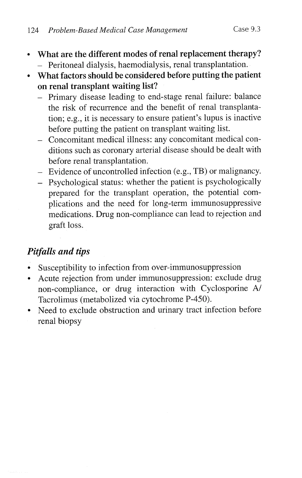
Examination case Nephrology 125
Examination case scenarios
i. This patient complained of malaise. Examine her
abdomen. (Transplanted kidney)
Important signs » Middle-aged woman with café-au-lait
complexion
e Arteriovenous fistula or graft;
or haemodialysis catheter (commonly
internal jugular vein)
e Parathyroidectomy scar on the neck
« Signs related to cyclosporin A usage
(e.g., gingival hyperplasia, hirsutism)
« Oblique left lower abdominal scar;
sometimes old Tenckhoff scars
¢ Oval dull mass in left or right lower
quadrant of abdomen
Diagnosis Transplant kidney for end stage renal
disease (graft failure)
Questions What are the common causes of end stage
renal failure in this age?
« Diabetic nephropathy, hypertensive
nephrosclerosis, lupus nephritis,
IgA nephropathy, focal segmental
glomerulosclerosis.
Pitfalls and tips » Do not miss Cushing’s features, fungal
infection, parathyroidectomy, and
nephrectomy scars.
View original page (figures / layout)
Page 10
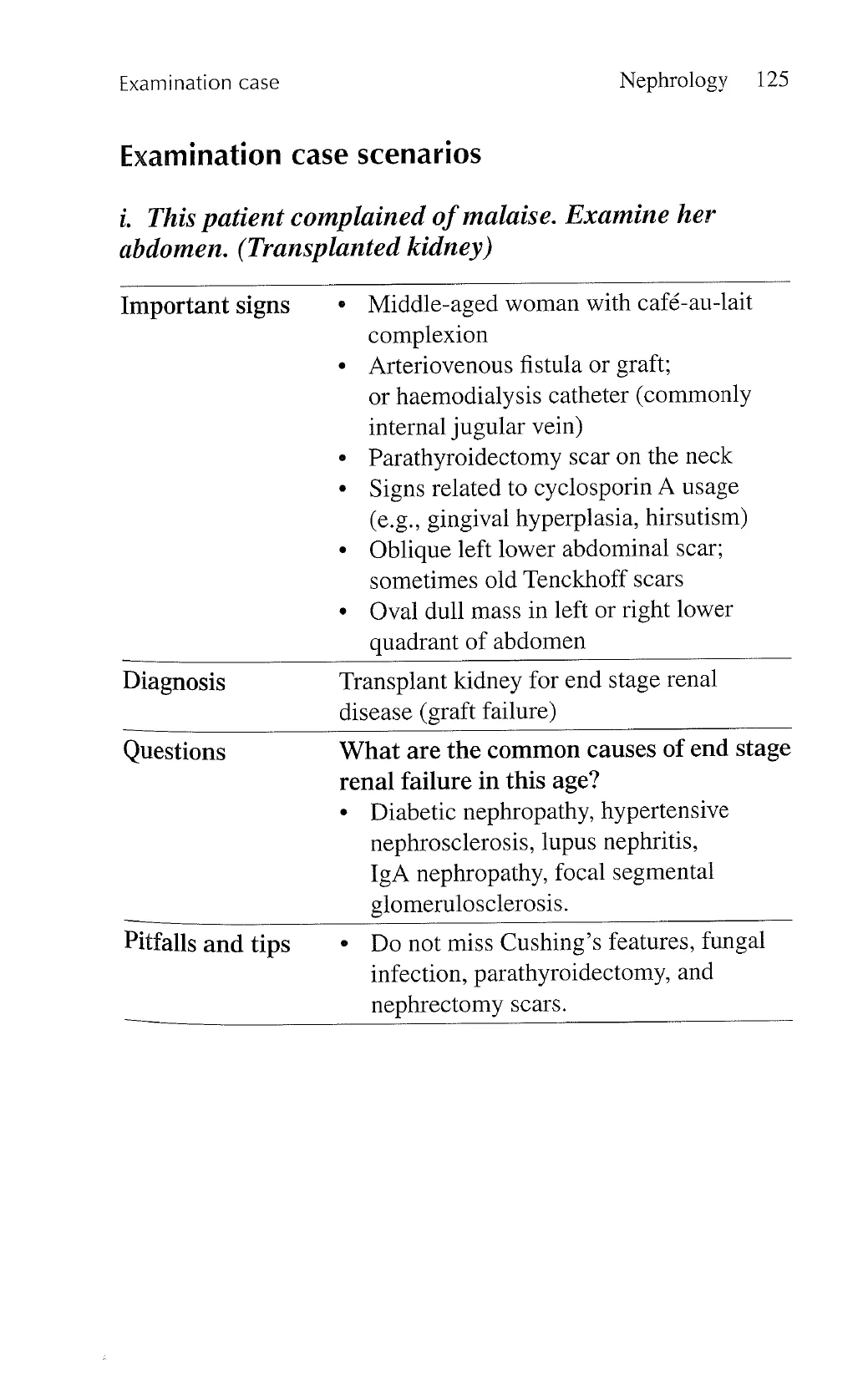
126 Problem-Based Medical Case Management Examination case
ii. Examine the abdomen (polycystic liver and kidney
disease on peritoneal dialysis).
Important signs .
Uraemic features
Tenckhoff catheter
Fluid inside the peritoneal cavity
Hepatomegaly with nodular surface
Bilateral ballotable kidneys
Diagnosis Polycystic kidney disease on peritoneal
dialysis
Pitfalls and tips .
Do not miss the liver.
View original page (figures / layout)